Intraabdominal Infections

Russell Lewis

2026-02-27

Intraabdominal Infections


Prof. Russell E. Lewis
Department of Molecular Medicine
University of Padua


russelledward.lewis@unipd.it
https://github.com/Russlewisbo


slides available at: www.padovaid.com

Learning objectives


  1. Classify intraabdominal infections (primary vs. secondary vs. tertiary)
  2. Understand epidemiology and risk factors for each infection type
  3. Master the microbiology and pathogenesis of IAI
  4. Apply diagnostic criteria for peritonitis variants
  5. Select appropriate antimicrobial therapy based on clinical context
  6. Recognize when source control is essential
  7. Manage CAPD-associated peritonitis
  8. Diagnose and treat intraperitoneal abscesses
  9. Risk-stratify patients for prognosis and outcomes
  10. Implement prevention strategies for secondary prophylaxis

Definition and classification

Peritonitis: Inflammatory response within the peritoneal cavity due to microbial or chemical contamination


By Location

  • Diffuse
  • Localized

By Type

  • Primary (spontaneous)
  • Secondary
  • Tertiary

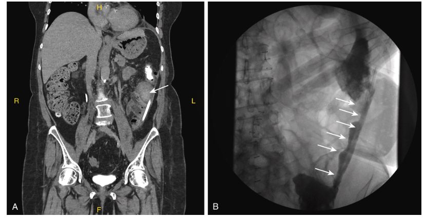
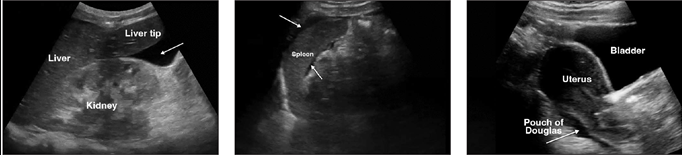
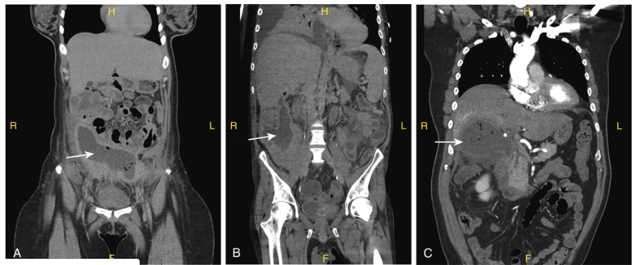
Primary vs. secondary vs. tertiary


Feature Primary Secondary Tertiary
Intraabdominal source None Present
(perforation, ischemia)
Persistent after
source control
Frequency ~1% of peritonitis cases 80–90% of cases Less common
Typical organisms Monomicrobial gram-negative Polymicrobial aerobic/anaerobic MDR, gram-negative, low virulence
Mortality 5–20% 10–40% 30–64%

Community-acquired vs. Healthcare-associated


Community-acquired (~80% of IAI)

  • Further subdivided: low-risk vs. high-risk
  • Risk factors: drug-resistant bacteria, severity (mild/moderate/severe), comorbidities
  • Common pathogens: E. coli, Klebsiella, anaerobes

Healthcare-associated

  • Complications of elective or emergency abdominal surgery
  • Nosocomial isolates (MRSA, MDR gram-negatives, Candida)
  • Higher mortality and morbidity

Uncomplicated vs. complicated infections


Uncomplicated IAI

  • Intramural inflammation of GI tract
  • Localized to single organ (e.g., appendicitis, cholecystitis)
  • No extension into peritoneal space

Complicated IAI (cIAI)

  • Extension beyond organ of origin
  • Peritonitis (diffuse or localized)
  • Abscess formation
  • Spillage into sterile peritoneal space

Primary peritonitis (SBP)

Primary peritonitis: Epidemiology


  • Prevalence — Accounts for ~1% of all peritonitis cases

    • Present in 10–30% of hospitalized cirrhotic patients with ascites
  • High-Risk Patient Groups

    • Cirrhosis (alcoholic, postnecrotic, cryptogenic)

    • Congestive heart failure

    • Nephrotic syndrome

    • Metastatic malignancy

    • Systemic lupus erythematosus

    • Rare in patients with no underlying disease

Risk factors for SBP development


  • High-volume ascites (advanced cirrhosis, Child-Pugh class C)

  • High Model for End-Stage Liver Disease (MELD) score

  • Coexisting gastrointestinal hemorrhage

  • Previous SBP episode

  • Low ascitic fluid protein (<1 g/dL)

  • Elevated serum bilirubin (>2.5 mg/dL)

  • Proton pump inhibitor use (reduces gastric acidity, increases translocation)


Primary peritonitis: Microbiology


Defining Characteristic: Monomicrobial infection

Most Common Organisms (in cirrhotic patients)

  1. Escherichia coli (most frequent)
  2. Klebsiella pneumoniae
  3. Other Enterobacterales
  4. Streptococcus pneumoniae (especially pediatrics)
  5. Other streptococci (including enterococci)
  6. Anaerobes (uncommon)
  7. Pseudomonas aeruginosa (rare)

Gram-Negative Dominance — 60–69% of cases caused by gram-negative enteric bacteria;
organisms presumed of gastrointestinal origin


Pathogenesis of primary peritonitis


Bacterial Translocation

  • Passage of viable bacteria from the GI tract lumen to mesenteric lymph nodes and bloodstream
  • Increased risk with portal hypertension and collateral vessel formation
  • Enhanced by decreased gastric acidity (PPI use) and altered bowel flora

Hematogenous Seeding

  • Bacteria reach peritoneal cavity via circulating blood
  • Explains predominantly monomicrobial pattern
  • Bacteremia present in 75% of cases with aerobic organisms

Host Factors

  • Reduced opsonic activity (low complement, reduced immunoglobulins)
  • Impaired polymorphonuclear cell function
  • Loss of anatomic barriers in advanced cirrhosis

Clinical presentation


  • Fever (> 37.8° C)

  • Nausea and vomiting

  • Diarrhea

  • Abdominal pain

    • Tenderness

    • Rebound tenderness

    • Bowel sounds hypoactive or absent

  • Cirrhosis - large volume ascites

Paracentesis


Diagnosis of SBP: Ascitic fluid examination

Essential Tests

  • WBC count with differential
  • Protein concentration
  • Gram stain
  • Culture (inoculate blood culture bottles)
  • Glucose
  • Amylase
  • Lactate dehydrogenase (LDH)
  • SAA (serum-ascitic albumin gradient)

Diagnostic Threshold

Parameter Significance
PMN >250/mm³ Presumptive SBP
Ascitic culture positive Confirms SBP
SAA >1.1 g/dL Suggests non-peritoneal ascites

SBP variants:
Monomicrobial nonneutrocytic bacterascites

Definition

  • Positive ascitic fluid culture
  • PMN <250 cells/mm³
  • No clinical peritonitis signs

Natural History

  • Resolves spontaneously in 62–86% of cases
  • Progresses to SBP in remainder (sometimes within hours)
  • Mortality same as classic SBP regardless of neutrophil response

Clinical Significance

  • May represent early colonization before host response
  • Symptomatic patients at higher risk for progression
  • Asymptomatic patients often remain colonized only
  • Treat if symptomatic or if organisms are gram-positive cocci

SBP variants:
Culture-negative neutrocytic ascites

Definition

  • PMN >250 cells/mm³
  • Negative ascitic fluid culture
  • No evident intraabdominal surgical source
  • Absence of pancreatitis

Epidemiology

  • Occurs in 35% of patients with clinical SBP
  • Blood culture positive in one-third of cases

Reasons for Culture Negativity

  • Antibiotic administration before sampling, Inadequate culture technique
  • Fastidious organisms

Improved Detection

  • Inoculate 10–20 mL ascitic fluid into blood culture bottles at bedside
  • Increases culture yield significantly

SBP variants:
Polymicrobial bacterascites

Definition

  • Multiple bacterial species on culture or Gram stain
  • PMN <250 cells/mm³
  • No elevation of ascitic protein

Etiology

  • Traumatic paracentesis (needle enters bowel)
  • Incidence: <1% of procedures
  • Risk factors: ileus, abdominal scars, adhesions

Natural History

  • Usually resolves spontaneously
  • Peritoneal fluid protein >1 g/dL and adequate opsonic activity predict spontaneous resolution
  • Treat only if symptomatic or if evolves to classic peritonitis

Clinical Pearl

Do not treat polymicrobial bacterascites empirically unless clinical deterioration occurs.

Treatment of SBP: Empiric therapy


First-Line Agent

  • Cefotaxime 2 g IV every 6–8 hours (or 3 g every 6 hours for high-risk patients) or other 3rd generation cephalosporin
  • Covers gram-negative enteric bacteria and streptococci
  • Can transition to oral after initial response
  • If recent history of MDR colonization or infection, may require broader-spectrum therapy

Alternative Regimens

Condition Agent
Non-severe Cefixime 400 mg PO BID
Renal impairment Adjust cephalosporin dosing
β-lactam allergy Fluoroquinolone (ciprofloxacin or norfloxacin)

Albumin supplementation

Indication

  • All patients with SBP
  • Particularly important in hemodynamically unstable patients

Albumin Dosing

  • 1.5 g/kg at diagnosis (maximum 100 g)
  • 1 g/kg on day 3

Mechanism

  • Expands plasma volume
  • Improves renal perfusion
  • Reduces variceal bleeding risk
  • Decreases renal dysfunction (hepatorenal syndrome)

Outcome Benefit

  • Reduces in-hospital mortality
  • Decreases kidney injury
  • Standard of care in SBP management

Treatment of SBP: Duration and outcomes


Duration of Therapy

  • 5 days of IV cefotaxime is sufficient for uncomplicated SBP (ineffective for ESBL producing isolates)
  • May transition to oral fluoroquinolone or cephalosporin
  • Repeat paracentesis not needed if clinical improvement occurs

Response Assessment

  • Clinical improvement (fever resolution, abdominal tenderness improves)
  • Laboratory response (declining WBC, improving renal function)

Prognosis After SBP

  • In-hospital mortality: 5–20%
  • Long-term survival: Poor—survivors warrant liver transplant assessment
  • Liver disease severity (MELD, bilirubin) predicts outcomes

Prevention of SBP: Primary prophylaxis

Indicated When

  • Low ascitic protein (<1.5 g/dL) +
    • Impaired renal function (creatinine >1.2 mg/dL) OR
    • Impaired liver function (bilirubin >2.5 mg/dL) OR
    • Low platelet count (<40,000/μL)

Agents for Primary Prophylaxis

Agent Dosing Notes
Norfloxacin 400 mg daily PO First-line
Ciprofloxacin 750 mg weekly PO Alternative
TMP-SMX 1 DS tablet daily Alternative

Efficacy

  • Reduces SBP incidence by 50–60%
  • Improves short-term survival
  • Cost-effective in high-risk patients
  • Recent meta-analysis have questioned efficacy

Prevention of SBP: Secondary prophylaxis

Indicated

  • All patients after first SBP episode
  • Reduces recurrence by 80%

Agents

  • Norfloxacin 400 mg daily (first-line)
  • Ciprofloxacin 750 mg weekly
  • TMP-SMX 1 DS tablet daily

Duration

  • Lifelong or until liver transplantation
  • Continue indefinitely to prevent recurrence

Additional Measures

  • Referral for liver transplant evaluation (poor prognosis after SBP)
  • Alcohol cessation counseling
  • Variceal screening and beta-blocker therapy

Secondary peritonitis

Secondary peritonitis: Epidemiology


Prevalence

  • Most common intraabdominal infection (80–90% of cases)
  • Results from visceral perforation or intraabdominal pathology

Common Causes

Surgical Emergencies

  • Perforated peptic ulcer
  • Ruptured appendicitis
  • Perforated diverticulitis
  • Acute cholecystitis/perforation

Other Sources

  • Ischemic bowel necrosis
  • Traumatic GI perforation
  • Gynecologic pathology (ruptured ovarian cyst, tubo-ovarian abscess)
  • Biliary or pancreatic disease

Etiologies of secondary peritonitis (representative)


Organ System Common Causes
Upper GI Perforated peptic ulcer, perforated gastric ulcer
Small bowel Meckel’s diverticulitis, small bowel perforation, ischemic necrosis
Appendix Perforated appendicitis
Colon Diverticulitis (perforation), toxic megacolon,
ischemic colitis
Biliary Perforation of gallbladder, cholangitis
Gynecologic Ruptured tubo-ovarian abscess,
perforated ovarian cyst
Trauma Iatrogenic or penetrating perforation

GI tract microbiota and bacterial density


Bacterial Population Gradient

  • Stomach: 10³–10⁴ CFU/mL (most anaerobes suppressed by acid)
  • Small intestine: 10⁴–10⁷ CFU/mL (increasing distally)
  • Terminal ileum: 10⁷–10⁹ CFU/mL (mixed aerobic/anaerobic)
  • Colon: 10¹¹–10¹² CFU/mL (predominantly anaerobes > aerobes by 1000:1)

Clinical Implication

  • Upper GI perforation: Fewer organisms, less anaerobic load
  • Lower GI perforation: Heavy polymicrobial inoculum with anaerobic dominance

Causes of secondary peritonitis

Organ Cause of Peritonitis
Distal esophagus Boerhaave syndrome
Malignancy
Trauma
Iatrogenic
Stomach Peptic ulcer perforation
Malignancy
Trauma
Iatrogenic
Duodenum Peptic ulcer perforation
Trauma
Iatrogenica
Biliary tract Cholecystitis
Stone perforation from gallbladder or common duct
Malignancy
Trauma
Iatrogenic
Pancreas Pancreatitis (e.g., alcohol, drugs, gallstones)
Trauma
Iatrogenic
Small bowel Ischemic bowel
Incarcerated hernia
Crohn disease
Malignancy
Meckel diverticulum
Trauma
Large bowel and appendix Ischemic bowel
Diverticulitis
Malignancy
Ulcerative colitis and Crohn disease
Appendicitis
Volvulus
Trauma (mostly penetrating)
Iatrogenic

Secondary peritonitis: Microbiology


Key Features

  • Polymicrobial (2–5 organisms typical, up to 10+)
  • Reflects normal GI flora
  • Aerobic-anaerobic polymicrobial common (not all may grown in culture)

Dominant Organisms

Category Examples Prevalence
Gram-negative rods E. coli, Klebsiella, Proteus, Enterobacter 60–80%
Anaerobes Bacteroides fragilis, anaerobic cocci, Clostridium 60–90%
Gram-positive cocci Streptococcus, Enterococcus, Staphylococcus 40–60%

Aerobic-anaerobic coverage in
secondary peritonitis

Early Infection (First Hours)

  • Gram-negative rod dominance (E. coli, Klebsiella)
  • Rapid multiplication and toxin production
  • Systemic toxemia and early sepsis
  • Clinical: fever, tachycardia, hypotension

Late Infection (Days)

  • Anaerobic bacteria proliferate (Bacteroides fragilis group)
  • Reduced oxygen tension favors anaerobic growth
  • Enhanced abscess formation (fibrin encapsulation)
  • Clinical: loculation, persistent fever despite initial therapy

Aerobic-anaerobic coverage in
secondary peritonitis, cont.

Clinical Consequences

  • Single-agent therapy (e.g., cephalosporin alone) inadequate
  • Requires dual coverage: aerobic + anaerobic agents
  • Source control essential to disrupt both populations

Pathogenesis and virulence factors in
secondary peritonitis

Initial Contamination

  • Spillage of GI flora into sterile peritoneal space
  • Magnitude of inoculum determines early severity
  • Bacterial adherence and LPS/endotoxin trigger inflammation

Host Response Activation

  • Complement activation (local and systemic)
  • Cytokine release (TNF-α, IL-1, IL-6, IL-8)
  • Polymorphonuclear recruitment to peritoneum
  • Increased vascular permeability

Bacterial Virulence Factors

  • Lipopolysaccharide (gram-negative endotoxin)
  • Capsule and fimbriae (adherence, invasion)
  • Toxins and enzymes (tissue invasion, abscess formation)
  • Antibiotic resistance (β-lactamase, efflux pumps)

Pathophysiology: Local response to
secondary peritonitis

Fibrin Deposition

  • Fibrinogen converts to fibrin clot at peritoneal surface, forms barrier limiting bacterial spread
  • Encapsulates contaminated area

Omental “Walling Off”

  • Greater omentum migrates to infection site, forms physical barrier around infection focus
  • Limits spread to distant peritoneal recesses

Peritoneal Fluid Exudation

  • Increased permeability → fluid accumulation containing antibodies, complement, white cells
  • Dilutes bacteria but may impair local immunity

Compartmentalization

  • Peritoneal reflections and mesenteric attachments route infection
  • Creates dependent recesses (pelvis, paracolic gutters, Morrison’s pouch)
  • Explains localization patterns and abscess locations


Pathophysiology: Systemic response to
secondary peritonitis


SIRS (Systemic Inflammatory Response Syndrome)

  • Fever, tachycardia, tachypnea, leukocytosis
  • Results from TNF-α, IL-1, IL-6 release
  • May progress to sepsis and multiorgan failure

Hemodynamic Changes

  • Initial phase: vasoconstriction (compensatory)
  • Late phase: vasodilation and increased capillary permeability
  • Hypovolemia and hypotension (septic shock)

Organ Dysfunction

  • Renal hypoperfusion → acute kidney injury
  • Pulmonary capillary leak → ARDS
  • Hepatic dysfunction → coagulopathy
  • GI hypomotility → ileus

Mortality Correlation

  • Extent of organ dysfunction predicts outcome
  • Reflected in APACHE II1 score, Mannheim Peritonitis Index2
  • Delayed recognition/treatment increases mortality

Clinical manifestations of secondary peritonitis

Symptoms

  • Acute abdominal pain (sudden onset if perforation)
  • Pain localized initially, generalizes with diffuse peritonitis
  • Nausea and vomiting (may be present or absent)
  • May have antecedent symptoms (dyspepsia before ulcer rupture, diarrhea before diverticulitis)

Physical Findings

  • Rebound tenderness and guarding (peritoneal irritation)
  • Absent or diminished bowel sounds (ileus)
  • Abdominal distention (third-spacing of fluid)
  • Hypotension and tachycardia (sepsis)
  • Fever (usually present but may be absent in elderly or immunocompromised)

Severity Indicators

  • Hemodynamic instability
  • Acute kidney injury
  • Leukocytosis >15,000 or left shift
  • Metabolic acidosis

Diagnostic workup: Laboratory studies


Complete Blood Count

  • WBC elevation (typically 12,000–20,000)
  • Left shift (immature bands) indicates severity
  • Absence of leukocytosis does not exclude peritonitis

Inflammatory Markers

Test Utility
C-reactive protein (CRP) Elevated; reflects severity
Procalcitonin >2 ng/mL suggests bacterial peritonitis; guides de-escalation
Lactate Elevated in sepsis; correlates with severity

Diagnostic workup: Laboratory studies, cont.


Chemistry

  • Renal function (creatinine, BUN)
  • Electrolytes (hypokalemia common from GI losses)
  • Liver function tests
  • Glucose (hyperglycemia or hypoglycemia possible)

Blood Cultures

  • Obtain before antibiotics if possible
  • Positive in 20–40% of secondary peritonitis
  • Guide specific organism coverage

Diagnostic imaging: CT abdomen/pelvis


Diagnostic imaging: CT performance


Sensitivity and Specificity

  • CT with IV contrast: 97% sensitivity for complicated IAI
  • Best imaging modality for secondary peritonitis and abscesses

Key CT Findings

  • Free air (pneumoperitoneum) → indicates perforation
  • Free fluid with inflammatory changes
  • Focal abscess collections
  • Source organ pathology (dilated colon, appendiceal thickening, etc.)
  • Evidence of ischemia (pneumatosis, portal venous gas)

Contraindications

  • Hemodynamic instability (may necessitate OR before imaging)

  • Renal insufficiency (contrast nephropathy risk)

  • Iodine allergy

Alternative imaging


  • Ultrasound: Portable, real-time, good for fluid assessment; lower sensitivity than CT
  • MRI: Excellent soft tissue detail; limited by cost and time; useful in renal insufficiency

Secondary peritonitis

Paracentesis and peritoneal cultures

Indications

  • Confirm diagnosis when imaging equivocal
  • Obtain organisms for culture and susceptibility
  • Obtain cell count and fluid analysis

Technique

  • Sterile procedure (surgical preparation)
  • Avoid areas of adhesion, stomas, surgical wounds
  • Obtain 20–30 mL in sterile container
  • Send for Gram stain, culture, cell count

Fluid Analysis

Parameter Finding
Appearance Turbid, purulent, bloody
PMN count Usually >50,000/mm³ in secondary peritonitis
Gram stain May show gram-negative rods, gram-positive cocci, anaerobes
Culture Polymicrobial growth expected

Clinical utility of paracentesis
in secondary peritonitis


  • Gram stain may guide initial therapy
  • Culture confirms organisms and guides de-escalation
  • Lower yield than in primary peritonitis (polymicrobial, fastidious anaerobes)

Prognosis:
Risk stratification in secondary peritonitis


APACHE II Score

  • Predicts mortality based on physiology, age, comorbidities
  • APACHE II >15 associated with 50% mortality
  • APACHE II >25 associated with >80% mortality

Mannheim Peritonitis Index (MPI)

Factor Points
Age >50 5
Female gender 5
Organ failure 7
Malignancy 4
Duration of peritonitis >24h 4
Origin (non-colonic) 4
Diffuse peritonitis 6
Exudate (purulent) 6


MPI Interpretation

  • MPI <21: 0% mortality

  • MPI 21–29: 11% mortality

  • MPI >29: 47% mortality

Tertiary peritonitis

Definition

  • Persistent or recurrent peritonitis despite successful source control of primary infection
  • Diagnosis: Peritonitis with signs of sepsis >48 hours after adequate surgery and source control

Epidemiology

  • Occurs in 3–10% of secondary peritonitis cases
  • Associated with delayed source control
  • Higher mortality: 30–64%

Microbiology

  • Less virulent organisms (coagulase-negative staphylococci, Candida)
  • Multidrug-resistant gram-negatives (Enterobacter, Pseudomonas)
  • MRSA
  • Often monomicrobial or sparse growth

Pathogenesis

  • Host immune dysfunction (exhaustion of cytokine response, impaired opsonization)
  • Biofilm-forming organisms
  • Inadequate source control or recurrent leak

Tertiary peritonitis: Management principles

Diagnostic Challenge

  • Distinguish from inadequately treated secondary peritonitis
  • Consider recurrent leak, anastomotic dehiscence, ischemia

Management Approach

  • Repeat imaging (CT) to identify new source
  • Selective repeat surgery only if surgically correctable source identified
  • Avoid routine re-exploration (may worsen outcomes)
  • Maximize supportive care: vasopressors, ECMO if needed, nutritional support
  • Broad-spectrum antimicrobials: carbapenem ± anti-Candida ± vancomycin pending cultures
  • Consider antifungal therapy (Candida common)

Prognosis

  • Mortality 30–64% despite appropriate management
  • Poor prognostic factors: organ failure, delayed recognition, immunosuppression
  • Consider goals of care discussion early

Antimicrobial therapy for
secondary peritonitis: An overview

Goal

  • Cover aerobes (gram-negative rods, gram-positive cocci) and anaerobes
  • Account for severity (mild-moderate vs. high-risk)
  • Consider prior antibiotic exposure (resistance risk)

Risk Stratification

Risk Category Features Typical Organisms
Low-risk Community-acquired, no recent hospitalization, no immunosuppression Susceptible gram-negatives, anaerobes
High-risk Healthcare-associated, recent surgery, immunosuppressed, prolonged hospitalization MDR gram-negatives, MRSA, Candida, enterococci

Timing

  • Initiate empiric therapy immediately (within 1 hour)
  • Source control should be initiated in parallel (not sequential)

Antimicrobial therapy for secondary peritonitis:
Drug classes for aerobic coverage

Beta-Lactams with beta-Lactamase Inhibitors

Agent Dosing Notes
Amoxicillin-clavulanate 875–125 mg TID Oral; limited spectrum
Ampicillin-sulbactam 3 g IV Q6H Good anaerobic coverage
Piperacillin-tazobactam 4.5 g IV Q6–8H Excellent coverage; pseudomonal

Cephalosporins (typically combined with metronidazole)

Agent Dosing Notes
Ceftriaxone 2 g IV Q12H +
metronidazole 500 mg TID
Lower pseudomonal coverage
Ceftazidime 2 g IV Q8H +
metronidazole 500 mg TID
Better Pseudomonas coverage

Carbapenems (broad spectrum, both Gram negative and anaerobes- reserve use)

  • Meropenem 1 g IV Q8H (or extended infusion)
  • Imipenem-cilastatin 500 mg IV Q6H
  • Ertapenem 1 g daily (does not cover Pseudomonas)
  • Excellent gram-negative and anaerobic coverage

Antimicrobial therapy for secondary peritonitis: Anaerobic coverage

Metronidazole

  • Dosing: 500 mg IV Q6–8H or 400–500 mg PO TID
  • Excellent anaerobic coverage
  • Minimal aerobic gram-negative coverage
  • Always combine with aerobic agent

Clindamycin

  • Dosing: 600 mg IV Q6–8H
  • Good anaerobic coverage
  • Some gram-positive aerobes covered
  • Emerging resistance in Bacteroides
  • Less preferred than metronidazole + cephalosporin

Carbapenems (cover both aerobes and anaerobes)

  • Single agent sufficient
  • Reserved for β-lactam-resistant organisms or severe disease

New antibiotics active against resistant
gram-negative bacilli


Agent ESBLs AmpC KPC OXA-48 MBL CRAB CRPA
Plazomicin
(not available in EU)
±
Eravacycline
Tigecycline
Temocillin
Cefiderocol
Ceftazidime/avibactam ±
Ceftolozane/tazobactam
Meropenem/vaborbactam
Imipenem/relebactam ±
Ampicillin/sulbactam + ceftazidime/avibactam ±

New agents for resistant organisms in
secondary peritonitis


Extended-Spectrum Agents

Agent Organism Coverage When to Use
Ceftolozane-tazobactam Pseudomonas, resistant GN Healthcare-associated, MDR risk
Ceftazidime-avibactam Carbapenem-resistant organisms, ESBLs Suspected resistance, prior carbapenems
Meropenem-vaborbactam Metallo-β-lactamases,
carbapenem-resistant
Last-resort therapy

Anti-Candida Agents


Agent Dosing When to Use
Fluconazole 400–800 mg/day Upper GI perforation, prolonged hospitalization
Anidulafungin 200 mg loading, then 100 mg/day Suspected azole-resistant Candida
Caspofungin 70 mg loading, then 50 mg/day Alternative to fluconazole, suspected azole-resistant Candida
Micafungin 100 mg daily Alternative to fluconazole, suspected azole-resistant Candida
Liposomal amphotericin B 3–5 mg/kg/day Suspected azole-resistant Candida, nephrotoxic

Vancomycin


  • Dosing: 15–20 mg/kg IV Q8–12H (goal trough 15–20 μg/mL)
  • For MRSA or severe penicillin allergy
  • Reserve use to avoid resistance

Source Control in Secondary Peritonitis


Principles of Source Control

  1. Identify and eliminate source (perforation, necrotic tissue, abscess)
  2. Operative debridement: Remove devitalized tissue, purulent material
  3. GI decompression: NG tube, venting catheter if ileus
  4. Peritoneal lavage: Saline irrigation to reduce bacterial load
  5. Drainage of dependent recesses (pelvis, paracolic gutters, Morrison’s pouch)
  6. Repair or resection of primary pathology

Timing

  • Emergent (within 1–2 hours): Perforation with peritonitis, uncontrolled sepsis
  • Urgent (within 6–12 hours): Contained abscess, clinical deterioration
  • Delayed (>24 hours): Selected patients with localized collection responding to antibiotics

Operative Techniques

  • Primary repair when possible (peptic ulcer perforation)
  • Resection with anastomosis vs. colostomy (depends on contamination, blood supply)
  • Avoid contamination of clean peritoneal surfaces

Supportive Care in Secondary Peritonitis

Hemodynamic Management

  • Aggressive fluid resuscitation (often requires 5–10 L in first 24 hours)
  • Vasopressors if hypotension persists after fluids (norepinephrine first-line)
  • Goal: Restore tissue perfusion, prevent organ failure

Respiratory Support

  • Mechanical ventilation if acute respiratory distress syndome (ARDS) develops
  • Positive end-expiratory pressure (PEEP) to improve oxygenation
  • Careful fluid management to balance resuscitation and pulmonary edema

Nutritional Support

  • Enteral nutrition when possible (preserves gut mucosa)
  • Total parenteral nutrition if unable to tolerate enteral feeds
  • Early nutrition improves outcomes and reduces infection risk

Organ Support

  • Renal replacement therapy if acute kidney injury develops
  • Correcting coagulopathy (fresh frozen plasma, vitamin K, platelets)
  • Managing hyperglycemia (insulin therapy, tight control)

Prevention of Postoperative Peritonitis

Preoperative Measures

  • Appropriate patient selection and optimization
  • Preoperative antibiotics (within 60 minutes of incision)
  • Hair clipping (not shaving) to prevent microabrasions

Intraoperative Measures

  • Strict aseptic technique
  • Avoid contamination during bowel manipulation
  • Gentle tissue handling to minimize ischemia
  • Adequate hemostasis
  • Maintain normothermia and normocapnia

Postoperative Measures

  • Remove drains/catheters as soon as possible
  • Monitor for signs of sepsis (fever, tachycardia, leukocytosis)
  • Early mobilization and feeding to promote GI function
  • Recognize anastomotic leaks early (CT imaging if deterioration)

Surgical Technique Factors

  • Primary vs. staged repair
  • Choice of anastomosis (mechanical, hand-sewn)
  • Drain placement (controversial; generally avoid unless high contamination)

Peritoneal-dialysis associated peritonitis

Peritoneal dialysis

PD-Associated peritonitis: Epidemiology

Incidence

  • Major complication of peritoneal dialysis (CAPD, APD)
  • Occurs in most dialysis patients over time
  • Recurrence rate: 20–30%

Primary Reason for Modality Failure

  • PD peritonitis recurrence is the leading cause of switch to hemodialysis
  • Repeated episodes increase peritoneal membrane damage

Risk Factors

  • Catheter-related factors: improper insertion, migration, tunnel infection
  • Touch contamination: improper bag exchange technique
  • Biofilm formation on catheter surface
  • Break in aseptic technique
  • Peritoneal membrane permeability changes
  • Patient-dependent factors: poor hygiene, younger age

PD Peritonitis: Microbiology

Organism Distribution

Category Prevalence Examples
Gram-positive 60–80% S. epidermidis, S. aureus, Streptococcus spp.
Gram-negative 15–30% E. coli, Klebsiella, Enterobacterales
Fungi 5–10% (increasing) Candida spp., rare molds
Mycobacteria <5% M. tuberculosis
(geographic dependent)

Common Pathogens

  • Coagulase-negative staphylococci (S. epidermidis): Most common gram-positive
  • S. aureus: Often from patient’s skin flora, more virulent
  • Streptococcus spp.: May indicate bowel translocation
  • Enterococci: Rare but significant if present
  • Pseudomonas: Often healthcare-associated, poor prognosis

Biofilm Considerations

  • S. epidermidis forms biofilm on catheter, protects from immune response and antibiotics

PD Peritonitis: Diagnosis

ISPD3 Criteria (International Society for PD)

Diagnosis requires 2 of 3:

  1. Cloudy peritoneal effluent (visual inspection)
  2. Peritoneal WBC >100 cells/mm³ (typically >50% PMN)
  3. Positive dialysate culture or Gram stain

Typical Laboratory Findings

Parameter Finding
Appearance Cloudy/turbid (vs. clear in health)
WBC count >100/mm³, usually 500–10,000
PMN predominance >50% (polymorphonuclear)
Bacteria Gram stain positive in 40–50%
Culture yield Positive in 90–95% with proper technique

Specimen Collection

  • Obtain specimen during fresh exchange
  • Use sterile technique
  • Send for cell count, differential, Gram stain, culture (aerobic and anaerobic)

PD Peritonitis: Treatment Principles

Empiric Therapy

  • Intraperitoneal (IP) antibiotics preferred
  • Cover gram-positives initially (cefazolin or vancomycin)
  • Add gram-negative coverage (ceftazidime or aminoglycoside)

Standard IP Regimens

Agent Dosing (Bolus / Maintenance) Target
Cefazolin 500 mg/2 L exchanges / 125 mg/2 L Gram-positive coverage
Ceftazidime 500 mg/2 L exchanges / 125 mg/2 L Gram-negative, Pseudomonas
Vancomycin 30 mg/kg loading / 15 mg/kg per exchange MRSA, resistant gram-positive

Duration

  • ISPD guidelines: 14–21 days of IP antibiotics, assess response at 2–5 days (peritoneal fluid should clear)
  • Culture results guide de-escalation

Dialysis catheter management


  • Continue CAPD during treatment (antibiotics via dialysate)
  • Remove catheter if no improvement by day 4–5
  • Remove catheter for fungal peritonitis (usually)
  • Remove if exit-site or tunnel infection present

PD Peritonitis: Fungal and Mycobacterial Infection

Fungal PD Peritonitis

  • Incidence: 5–10% of all CAPD peritonitis: Most common: Candida spp. (>90% of fungal cases)
  • Risk factors: prior antibiotics, immunosuppression, diabetes

Management of Fungal Peritonitis

  • Antifungal agents (fluconazole, amphotericin B): Often requires IV therapy in addition to IP
  • Mandatory catheter removal (biofilm barrier limits drug penetration)

Mycobacterial PD Peritonitis

  • M. tuberculosis: Geographic variation (high in endemic areas)
  • Nontuberculous mycobacteria (NTM): Rising incidence

Management of Mycobacterial Peritonitis

  • Long course of anti-TB drugs (6+ months for TB)
  • Continue CAPD during treatment (if possible)
  • Catheter removal for persistent infection (NTM)
  • Diagnosis delay common (insidious presentation)

PD Peritonitis: Outcomes and Catheter Removal

Outcomes with Treatment

  • Resolution: 90–95% of bacterial peritonitis cured with appropriate therapy
  • Recurrence: 20–30% of patients experience repeat episodes
  • Modality failure: 30–40% eventually switch to hemodialysis

Indications for Catheter Removal

  • Fungal peritonitis (almost universally)
  • Failure to respond by 4–5 days of therapy
  • Exit-site or tunnel infection with peritonitis
  • Polymicrobial peritonitis (suggests bowel perforation; need imaging)
  • Refractory peritonitis (multiple episodes with same organism)
  • Patient choice (prefer hemodialysis)

Prevention of Recurrence

  • Patient education (proper exchange technique, hand hygiene)
  • Catheter modification (Y-set or disconnect systems)
  • Use of prophylactic topical antibiotics (mupirocin)
  • More frequent exchanges during acute peritonitis
  • Prompt treatment of exit-site infections

Intraperitoneal Abscess

Intraperitoneal Abscesses: Definition and Epidemiology

Definition

  • Focal collections of pus within the peritoneal cavity
  • Walled-off by fibrin, omentum, and peritoneum
  • Complications of primary or secondary peritonitis

Common Locations

Location Frequency Clinical Features
Pelvic 30–40% Dependent site; difficult to examine
Paracolic gutters 20–30% Follow colon anatomy
Subphrenic 15–20% Upper abdomen; may irritate diaphragm
Perihepatic 10–15% Near hepatic hilum, fissures
Morrison’s pouch 5–10% Dependent upper abdomen

Formation Mechanism

  • Peritoneal routes of drainage determine abscess site
  • Right paracolic gutter → pelvis (gravity)
  • Upper abdomen → subphrenic spaces
  • Lesser sac collections from anterior abdominal pathology

Intraperitoneal Abscesses: Microbiology

Organism Characteristics

  • Polymicrobial (usually 2–5 organisms)
  • Reflects organisms from primary source (GI tract)
  • Similar to secondary peritonitis microbiology

Common Organisms

  • *E. coli* (gram-negative rod)
  • Bacteroides fragilis (obligate anaerobe)
  • Streptococcus spp. (gram-positive cocci)
  • Enterococcus spp. (gram-positive coccus)
  • Other anaerobes (Peptostreptococcus, Clostridium)

Culture Characteristics

  • Lower culture yield than peritoneal fluid (bacteria within abscess wall)
  • Gram stain may guide initial therapy
  • Anaerobic cultures essential (often missed if not specifically requested)

Implications for Therapy

  • Broad-spectrum empiric coverage needed
  • β-lactam + anaerobic agent, or carbapenem
  • Culture-guided de-escalation critical
  • Drainage required in addition to antibiotics

Intraperitoneal Abscesses: Clinical Presentation

Symptoms - Fever (may be low-grade, intermittent) - Localized abdominal or flank pain (depending on location) - GI symptoms: nausea, vomiting, anorexia (if gastric irritation) - Urinary symptoms: dysuria, frequency (if bladder compression) - Subphrenic abscess: Shoulder pain (referred), dyspnea

Physical Findings - Localized abdominal tenderness (over abscess site) - Palpable mass (large collections only) - Fever - Hemodynamic instability (if large, with systemic toxicity)

Laboratory/Imaging Abnormalities - Leukocytosis (may be modest in chronic abscess) - Elevated CRP - CT findings: Fluid collection with enhancing rim, air-fluid level if gas-forming organism

Intraperitoneal Abscesses: Diagnosis

Imaging Modalities

Modality Sensitivity Utility
CT with IV contrast 95–97% Gold standard; localization; guidance for drainage
Ultrasound 90–95% Real-time, portable; bedside assessment
MRI 90–95% Excellent soft tissue; limited in acute care

CT Findings

  • Fluid collection with enhancing rim
  • Air-fluid level (gas-forming organisms)
  • Loculation (multiloculated abscesses)
  • Associated organ pathology
  • Size and location relative to surrounding structures

Paracentesis (if not already done)

  • May be diagnostic if fluid obtained
  • Often done as precursor to drainage
  • Culture crucial for organism identification

CT imaging: intraperitoneal abscess

Liver abscess

Intraperitoneal Abscesses: Treatment — Drainage Decisions

Percutaneous vs. Surgical Drainage

Factor Percutaneous Surgical
Access Image-guided (CT/US) Direct visualization
Invasiveness Minimally invasive Formal operation
Morbidity Lower Higher (general anesthesia, incisions)
Efficacy 80–90% success Near 100% success
Best used in Accessible collections, stable patient Inaccessible collections, unstable patient

Selection Criteria for Percutaneous Drainage

  • Accessible collection (not multiloculated or complex)

  • 3–4 cm in diameter (smaller often respond to antibiotics alone)

  • Hemodynamically stable patient

  • Expertise available for image-guided drainage

When Surgery is Preferred

  • Inaccessible collections (multiloculated, complex)
  • Failure of percutaneous drainage
  • Hemodynamic instability
  • Associated pathology requiring intervention

Intraperitoneal Abscesses: Antibiotic Therapy

Duration and Route

  • IV therapy initially (acute abscess)
  • Transition to oral after clinical improvement (usually 7–10 days IV, then oral)
  • Total course: 10–14 days (shorter if source controlled and drained)

Empiric Regimen

  • β-lactam + β-lactamase inhibitor: Piperacillin-tazobactam 4.5 g Q6–8H
  • Or cephalosporin + metronidazole: Ceftriaxone 2 g Q12H + metronidazole 500 mg TID
  • Or carbapenem: Meropenem 1 g Q8H

Transition to Oral

  • After clinical improvement and drain output minimal
  • Options: Amoxicillin-clavulanate, fluoroquinolone + metronidazole (limited)

Culture-Directed De-escalation

  • Narrow spectrum once organism susceptibilities known
  • Select oral agent based on susceptibilities and drug properties
  • Complete course based on clinical response (not arbitrary duration)

Intraperitoneal Abscesses: Prognosis

Outcomes with Appropriate Management

  • Cure rate: 85–95% with combined drainage and antibiotics
  • Mortality: 5–15% (higher in elderly, immunocompromised)

Factors Affecting Prognosis

Factor Impact
Time to diagnosis Delayed diagnosis → higher mortality
Size >5 cm Larger collections → worse prognosis
Multiple organisms (esp. anaerobes) More virulent; worse outcome
Associated peritonitis Indicates severe primary disease
Patient age, comorbidities Older, immunocompromised → worse

Complications of Treatment

  • Recurrent abscess: May occur if inadequate drainage or source not controlled
  • Fistula formation: If drain erodes into viscus
  • Drain site infection: May progress if not managed
  • Incomplete resolution: Imaging follow-up recommended at 4–6 weeks

SECTION 5: SUMMARY AND KEY TAKEAWAYS

Clinical Pearl: The 10 Key Takeaways on IAI

  1. Primary peritonitis (~1% of cases) occurs without evident abdominal source; typically monomicrobial gram-negatives in cirrhotic patients; diagnosis confirmed by PMN >250/mm³ and positive culture.

  2. SBP prevention with antibiotics (norfloxacin primary prophylaxis) reduces incidence 50–60%; secondary prophylaxis after first episode is lifelong.

  3. Secondary peritonitis (80–90% of cases) is polymicrobial, requires source control, and mortality is more dependent on adequacy of drainage/surgery than antibiotic choice.

  4. Empiric therapy must cover aerobes and anaerobes; β-lactam + β-lactamase inhibitor or cephalosporin + metronidazole are standard options for community-acquired secondary peritonitis.

  5. Tertiary peritonitis (persistent after source control) carries 30–64% mortality; repeated surgery often worsens outcomes; supportive care and selective re-imaging are key.

Clinical Pearl: Key Takeaways (continued)

  1. CT imaging is 97% sensitive for complicated IAI; pneumoperitoneum indicates perforation and need for urgent surgery.

  2. Fever is not always present in elderly, immunocompromised, or on immunosuppressive therapy; maintain high suspicion with abdominal pain and leukocytosis.

  3. CAPD peritonitis is predominantly gram-positive (touch contamination) and responds to IP antibiotics in 90–95%; fungal peritonitis requires catheter removal.

  4. Intraperitoneal abscesses require both drainage and antibiotics; percutaneous drainage under imaging guidance is now first-line for accessible, hemodynamically stable patients.

  5. Early recognition and source control are critical to outcome; every hour of delay in antibiotics increases mortality; source control (surgery or drainage) should proceed in parallel with antimicrobials, not sequentially.

Algorithm: Approach to Suspected IAI

When to Worry: Red Flags for MDR Organisms


High-Risk Features for Resistant Pathogens

  • Healthcare-associated infection (recent hospitalization, surgery, invasive procedure)
  • MRSA colonization or prior MRSA infection
  • Prior broad-spectrum antibiotics (β-lactams, carbapenems, fluoroquinolones)
  • Immunosuppression (chemotherapy, transplant, HIV with CD4 <200)
  • Severe illness at presentation (septic shock, APACHE >25)
  • Candida species in blood cultures or peritoneal fluid
  • Diabetic or renal failure patients
  • Polymicrobial bacteremia with unusual organisms

Modified Empiric Therapy for High-Risk Patients

  • Extended-spectrum agent: Ceftazidime-avibactam or ceftolozane-tazobactam
  • Add vancomycin for MRSA coverage
  • Add antifungal (fluconazole or echinocandin) if high risk for Candida
  • Consider consulting infectious diseases specialist

Quick Reference:
Empiric Antibiotic Selection by Scenario


Clinical Scenario First-Line Empiric Alternative Duration
Primary peritonitis (SBP) Cefotaxime 2 g Q6–8H Ciprofloxacin 400 mg PO Q12H 5 days
Secondary peritonitis, CA Piperacillin-tazobactam 4.5 g Q6–8H Ceftriaxone 2 g Q12H + metronidazole 500 mg TID 10–14 days
Secondary peritonitis, HA 3rd-4th gen Cephalosporin + vancomycin + anti-Candida Carbapenem ± antifungal 10–14 days
CAPD peritonitis Cefazolin IP + ceftazidime IP Vancomycin IP 14–21 days
Intraperitoneal abscess Piperacillin-tazobactam 4.5 g Q6–8H Ceftriaxone + metronidazole 10–14 days

Abbreviations: CA = community-acquired; HA = healthcare-associated; IP = intraperitoneal

Further Learning

Key References

  • Solomkin JS, et al. Infectious Diseases Society of America guidelines for complicated intra-abdominal infections. Clin Infect Dis. 2017;64(7):e69-e89.

  • Carbapenem-resistant Enterobacteriaceae: Emerging threat and options for infection control. Am J Infect Control. 2012.

  • Multidrug-resistant organisms and healthcare epidemiology: Recent advances and clinical perspectives. Crit Care Med. 2023.

References


1.
Hodzic S Delibegovic S Markovic D. Apache II scoring system is superior in the prediction of the outcome in critically ill patients with perforative peritonitis.  med arh. Vol. 65. 2011;65(176):82–5.
2.
Jain S Sharma R Ranjan G. A prospective study evaluating utility of mannheim peritonitis index in predicting prognosis of perforation peritonitis.  j nat sci biol med. 6(suppl 1). 2015;6(suppl 1)(178):S49–52.
3.
Cho Y Li PK-T Chow KM. ISPD peritonitis guideline recommendations: 2022 update on prevention and treatment.  perit dial int. Vol. 42. 2022;42(364):110–53.