Enteric Infections-Infectious diarrhea

Russell E. Lewis

2026-03-01

Enteric Infections- Infectious Diarrhea


Prof. Russell E. Lewis
Department of Molecular Medicine
University of Padua


russelledward.lewis@unipd.it
https://github.com/Russlewisbo


slides available at: www.padovaid.com

Learning objectives


  • Identify common causes of infectious diarrhea in adults in developed countries
  • Describe patient history and clinical presentation distinguishing viral vs. bacterial causes
  • Recognize warning signs for severe diarrheal disease
  • Describe management approach and treatment

Overview — Global burden


  • Infectious diarrhea: top 10 cause of death worldwide (Troeger et al., 2018)
  • 1.7 billion cases annually
  • Leading cause of death in children under 5 years (Liu et al., 2016)
  • In adults in resource-rich settings: often “nuisance disease” with key clinical decision points

Definitions and duration


Diarrhea

  • Passage of loose or watery stools

  • ≥3 times in 24 hours

  • Abnormal stool frequency or consistency

Duration Categories

  • Acute: <14 days

  • Persistent: 14-30 days

  • Chronic: >30 days


Dysentery: diarrhea with visible blood, associated with fever and abdominal pain

Pathophysiology of diarrhea


Normal intestinal physiology

  • GI tract absorbs 8-9 L fluid daily

  • Net secretion only 100-200 mL/day

  • Pathogen virulence factors disrupt this balance

Three mechanisms of pathogen damage

  • Altered ion absorption/secretion

  • Disruption of epithelial barrier

  • Villus atrophy and enzyme deficiency

Small bowel vs. large bowel diarrhea


Small Bowel Pattern

  • Large volume stools (>200 mL/stool)

  • Watery consistency

  • Cramping periumbilical pain - 4-8 stools daily

Associated Symptoms: Nausea/vomiting common, weight loss possible

Large Bowel Pattern

  • Small volume stools (<200 mL/stool)

  • Frequent passage (>5-6/day)

  • Painful tenesmus and urgency

  • Bloody or mucoid stools

Associated Symptoms - Abdominal cramping/pain -sytemic symptoms less common

Overview of infectious etiologies

  • Most diarrhea is viral: stool cultures positive only 1.5-5.6%
  • Viral: norovirus (most common), rotavirus, adenoviruses 40/41, astrovirus
  • Bacterial: Salmonella, Campylobacter, Shigella, ETEC, EHEC/STEC
  • Parasitic: Cryptosporidium, Giardia, Cyclospora, Entamoeba

Norovirus — “The winter vomiting virus”


Key Features

  • Most common cause of acute gastroenteritis worldwide (Ahmed et al., 2014)

  • Affects all ages, including highly immune populations

  • Mean incubation: 24-48 hours - “Winter vomiting disease” (Northern hemisphere)

Norovirus epidemiology & transmission

Viral Characteristics

  • Non-enveloped RNA virus, Caliciviridae family - Multiple genotypes; no lasting immunity after infection (Patel et al., 2008)

  • Extremely stable: resists alcohol, chlorine, temperatures to 60°C

Transmission Routes

  • Primarily fecal-oral >aerosol transmission documented

  • Fomite transmission (contaminated surfaces)- Can survive environmental conditions for weeks
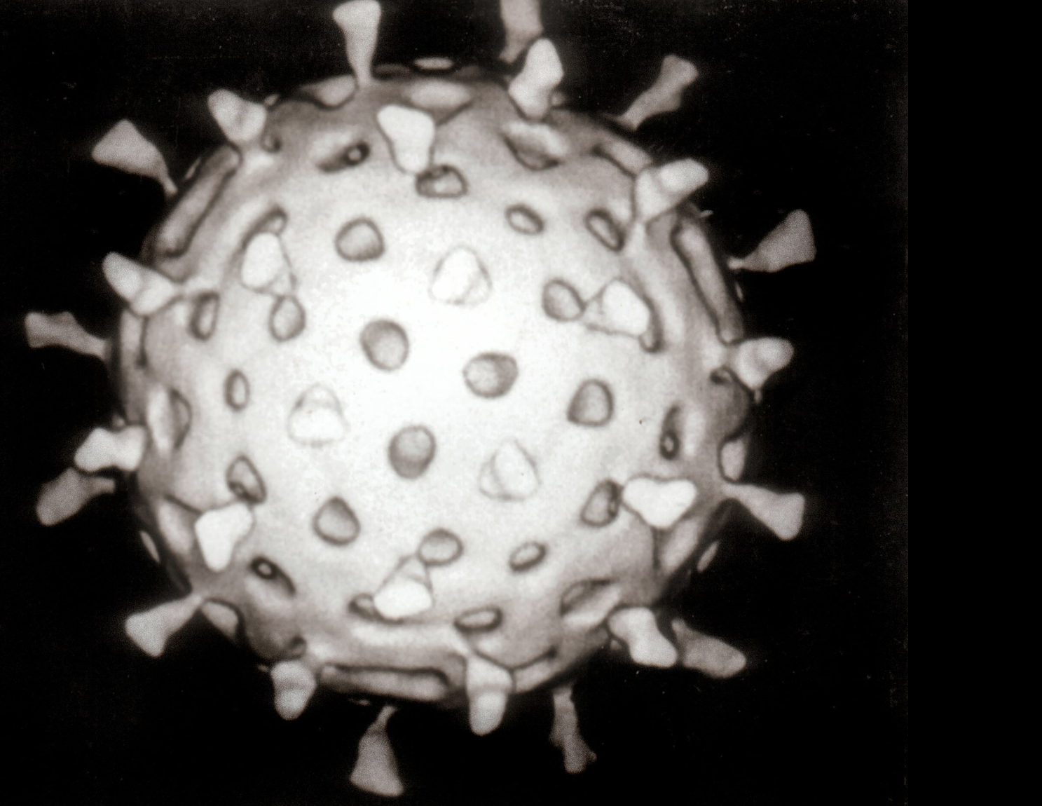
Norovirus clinical manifestations


Symptoms

  • Acute onset vomiting (prominent feature)

  • Watery non-bloody diarrhea (4-8 stools/24 hours)

  • Fever in 50% of cases

  • Malaise and headache

Clinical Course

Duration typically 48-72 hours

Complete resolution expected

Dehydration is main complication

Secondary bacterial infection rare

Diagnosis & Management: Clinical diagnosis in outbreak setting - EIA or PCR for confirmation (primarily epidemiologic)

Treatment: supportive care and oral rehydration solution

Norovirus in immunocompromised patients


Unique Clinical Course

  • Chronic infection: shedding for months to years

  • Viral evolution occurs during infection

  • Severe, refractory symptoms possible

  • May lead to malnutrition and functional decline

Treatment Challenges

  • No specific antiviral therapy proven effective

  • Supportive care remains cornerstone

  • Probiotic therapy: insufficient evidence

  • Management: supportive nutrition, hydration

Norovirus outbreak management


Prevention Measures

  • Hand hygiene with soap and water (alcohol ineffective)

  • Environmental cleaning with chlorine-based disinfectants (0.5-1% bleach)

  • Surface decontamination: quaternary ammonium compounds

  • Isolation precautions for symptomatic patients

Outbreak Control

  • Early detection and reporting to public health

  • Exclusion of food handlers until 48 hours symptom-free

  • Restriction of admitted patients in healthcare settings

Rotavirus overview


Epidemiology

  • Most common cause of severe diarrhea in children worldwide (Parashar et al., 2006) - >100 million cases annually

  • Approximately 150,000 deaths in children <5 years (Tate et al., 2016)

  • Peak incidence: 6-24 months age

Clinical Course

  • Duration 3-8 days , often more severe than norovirus

  • Dehydration: primary complication

Rotavirus pathophysiology

Viral Characteristics

  • 70 nm non-enveloped RNA virus (Reoviridae family)

  • Segmented genome with multiple genes encoding virulence factors

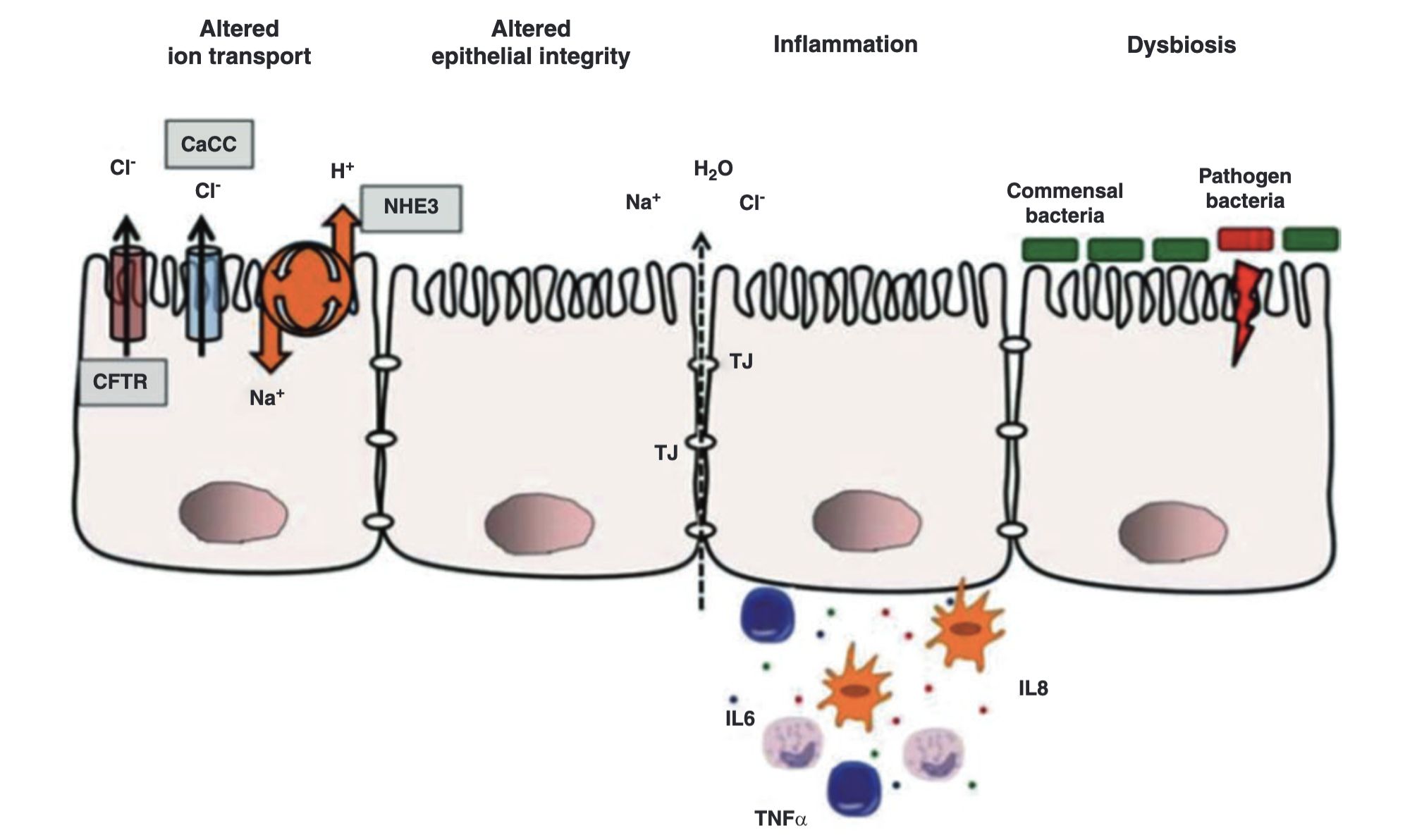
Mechanisms of Diarrhea - Villus shortening and disruption - Brush-border enzyme deficiency (lactase, sucrase) - Calcium-dependent enterotoxin production (NSP4) - Impaired water and ion absorption

Rotavirus vaccines


Available Vaccines

RotaTeq (Pentavalent) - Manufactured by Merck - 3-dose series - RV1, RV2, RV3, RV4, RV5

Rotarix (Monovalent) - Manufactured by GSK - 2-dose series - RV1 genotype coverage

Impact on Disease:

Rotavirus — Key clinical points


Typical presentation

  • Watery, non-bloody diarrhea

  • Vomiting less prominent than with norovirus

  • Respiratory symptoms occasionally present (suggests dual viral infection)

Risk factors for severe disease

  • Age <24 months

  • Malnutrition

  • Lack of prior exposure/vaccination

  • Comorbid conditions

Epidemiology

  • Common in daycare settings and seasonal: winter months and dry seasons in temperate climates

Other viral pathogens


Virus Age Group Key Features
Sapovirus Children Similar to norovirus; outbreaks
Astrovirus Young children Milder than rotavirus
Adenovirus 40/41 Infants/toddlers Winter seasonality
Enteroviruses Variable Rash sometimes present
Coronaviruses
(SARS-CoV-2)
All ages Mild GI symptoms often with respiratory

Bacterial etiologies

Enterotoxigenic E. coli (ETEC) overview

Epidemiology & Pathogenesis

  • Leading cause of acute diarrhea in developing countries (Qadri et al., 2005)

  • Survives in water; transmitted via contaminated food/water

  • Produces enterotoxins: heat-labile (LT) and heat-stable (ST) toxins

Clinical Presentation

  • Watery diarrhea, often dehydrating

  • Nausea common; vomiting less frequent

  • Fever absent or mild

  • Duration typically 3-5 days
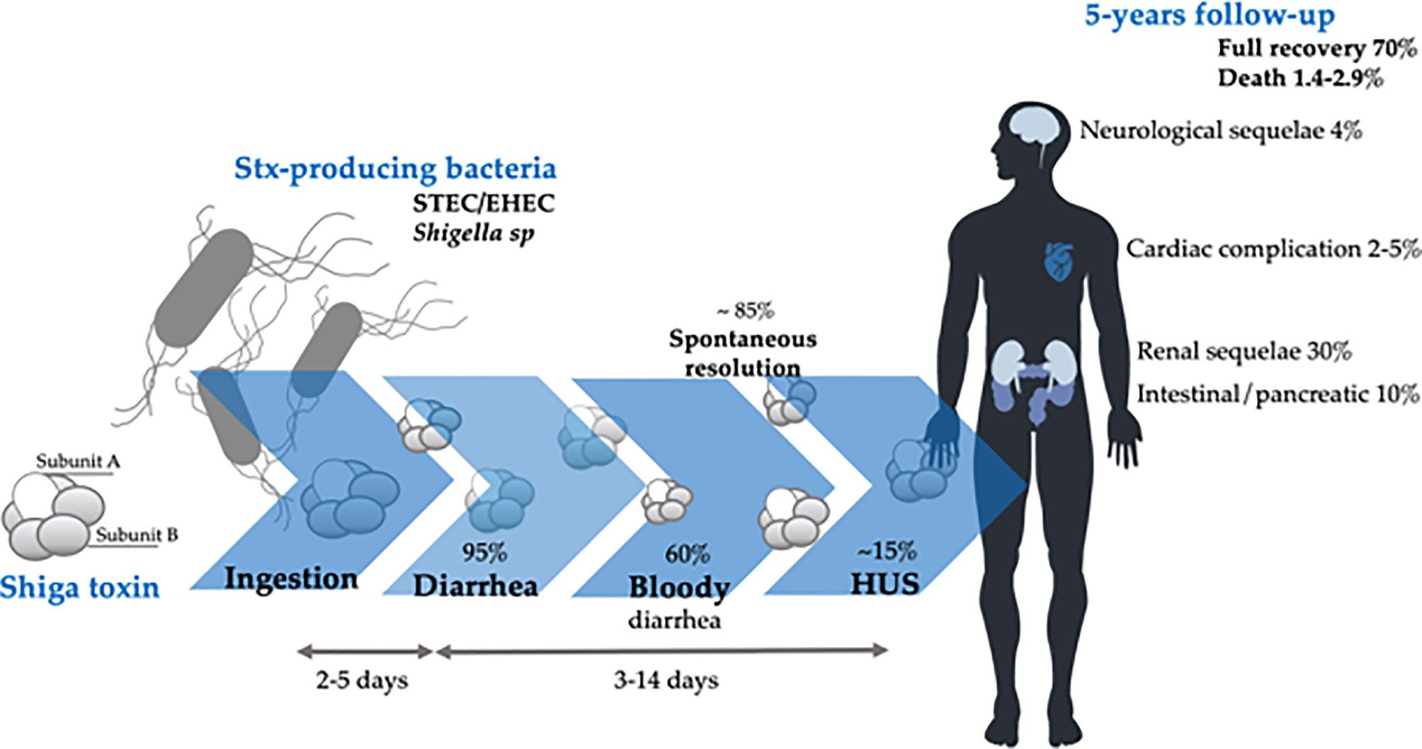
ETEC Toxins and mechanisms


Heat-Labile Toxin (LT)

  • Similar to cholera toxin

  • Activates adenylate cyclase

  • Increases cAMP

  • Stimulates secretion

Heat-Stable Toxin (ST)

  • Smaller molecular weight

  • Activates guanylate cyclase

  • Increases cGMP

  • More tissue-specific

Result: Increased intestinal cyclic nucleotides → electrolyte and water secretion → watery diarrhea

Other Pathogenic E. coli Strains


EPEC (Enteropathogenic E. coli)

  • Primarily affects children <6 months

  • Contains Eae gene encoding adhesin

  • Causes attaching and effacing lesions - Non-bloody watery diarrhea

EIEC (Enteroinvasive E. coli)

  • Invasive mechanism similar to Shigella

  • Bloody diarrhea with systemic symptoms - Fever and abdominal pain common

EAEC (Enteroaggregative E. coli)

  • Biofilm formation on epithelium- causes persistent or chronic non-bloody diarrhea (>14 days)

  • Often associated with travel to developing countries

EHEC/STEC overview


Clinical Significance

  • Shiga toxin-producing E. coli (STEC) strains

  • E. coli O157:H7 most common in North America (Karch et al., 2005)

  • Multiple non-motile serotypes cause disease

  • Shiga toxin causes microangiopathic hemolytic damage (Tarr et al., 2005)

Hemolytic Uremic Syndrome (HUS)


STEC-Associated HUS

  • Occurs in approximately 5-15% of STEC infections

  • Often follows 3-5 days of hemorrhagic diarrhea

  • Triad: microangiopathic hemolytic anemia (schistocytes on blood smear), thrombocytopenia, acute kidney injury

Prognosis and Sequelae

  • 5-year outcomes: ~70% complete recovery Mortality: 1.4-2.9%

  • Chronic sequelae: renal dysfunction (8-50%), neurological (5-25%), cardiac (5%)

HUS management


Critical Principle: Avoid Antibiotics !!!

  • Antibiotic use associated with 25% increase in HUS risk

  • Proposed mechanism: bacterial lysis releases Shiga toxin

  • Even fluoroquinolones and azithromycin increase risk

  • Avoid antimotility agents

HUS Management

Renal replacement therapy: essential in ~50% of cases

Blood product support: transfusions for anemia, platelets carefully

Plasma exchange: controversial but may help neurologic complications

ICU-level supportive care often required

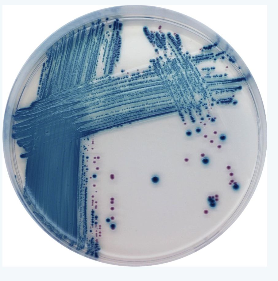
EHEC Detection Methods

Diagnostic Approaches

  • Sorbitol MacConkey agar: STEC O157:H7 appears non-sorbitol fermenting (colorless)

  • Chromogenic agar: substrate produces color with specific enzymes

  • EIA for Shiga toxins: rapid detection from stool

  • PCR for Stx genes: confirmatory molecular testing

Chromogenic agar: STEC strains grow as mauve colonies, while other bacteria grow as blue, colorless, or are inhibited

Chromogenic agar: STEC strains grow as mauve colonies, while other bacteria grow as blue, colorless, or are inhibited

EHEC Outbreaks — Germany 2011


2011 Outbreak Details

  • Strain: O104:H4 (unusual non-motile strain) (Rasko et al., 2011)

  • Total infected: 12,600 cases

  • HUS cases: 4,321

  • Deaths: 50 (42 from HUS, 8 from sepsis)

Unique Features

  • Prophage carrying Stx gene plus additional virulence genes

  • Multidrug resistance including fluoroquinolone resistance

  • Foodborne outbreak traced to sprouts from Egypt

  • Deadliest STEC outbreak in modern history
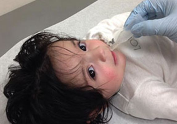
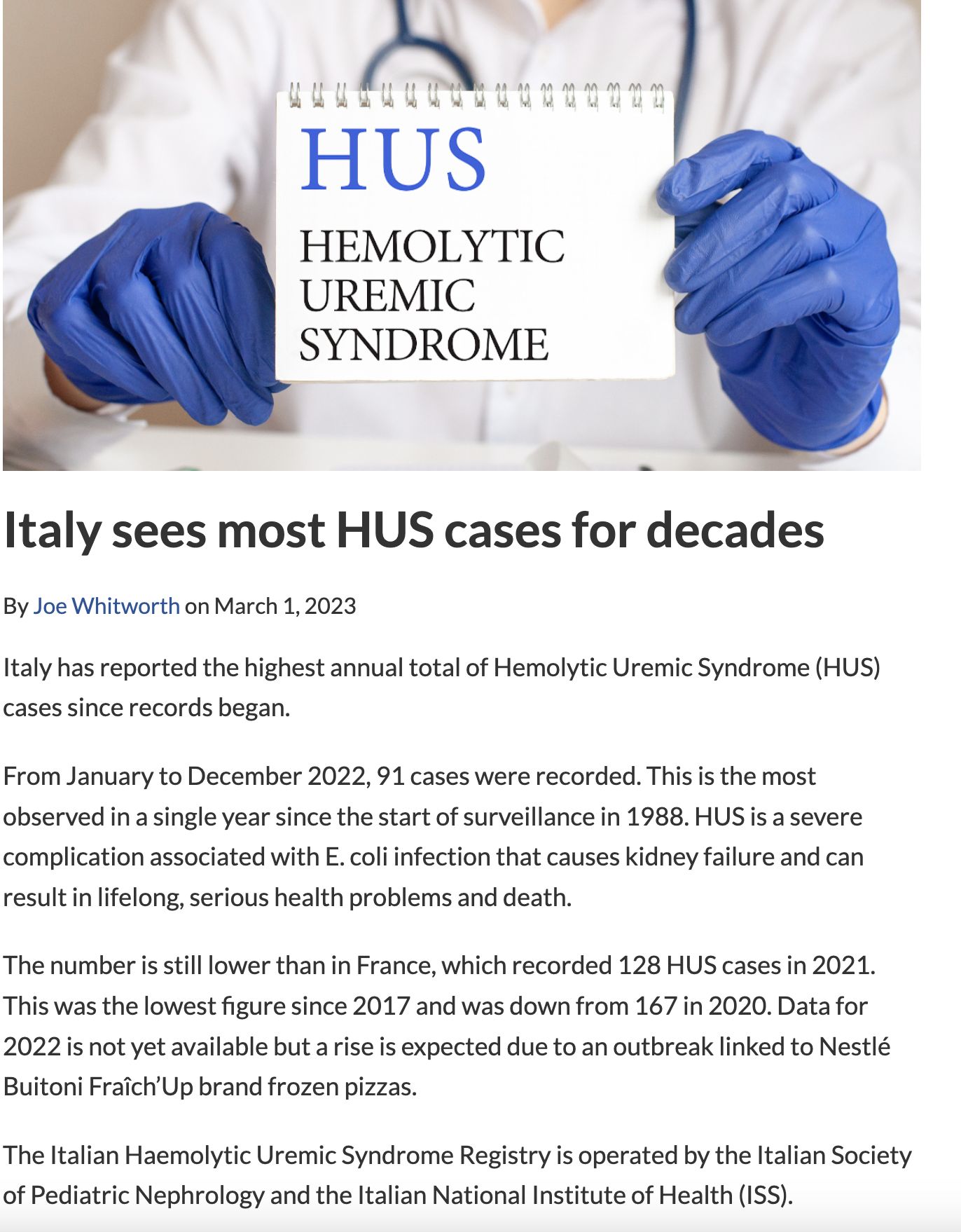
HUS in Italy?

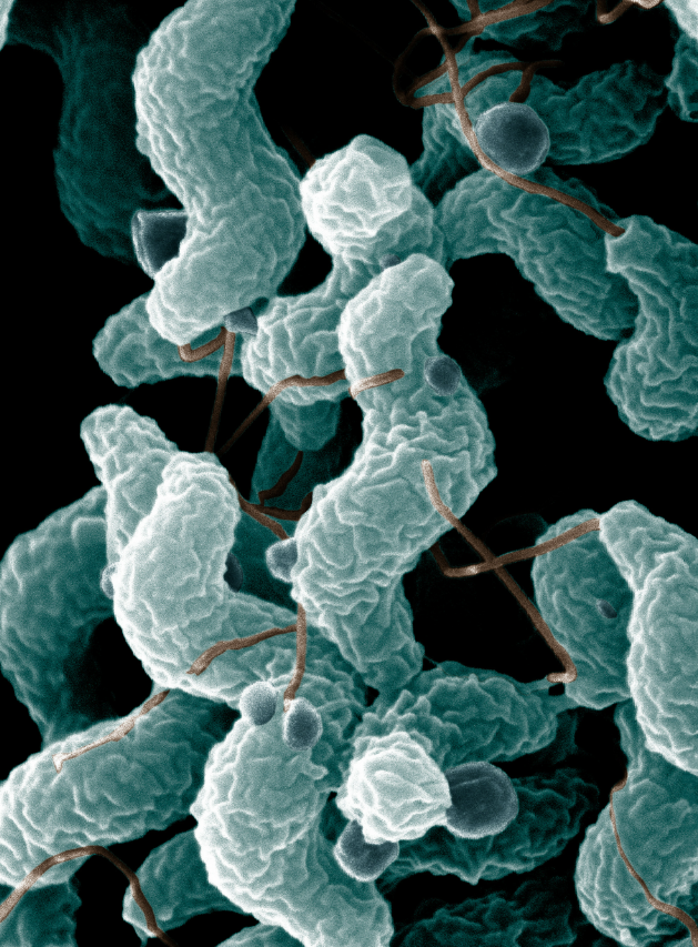
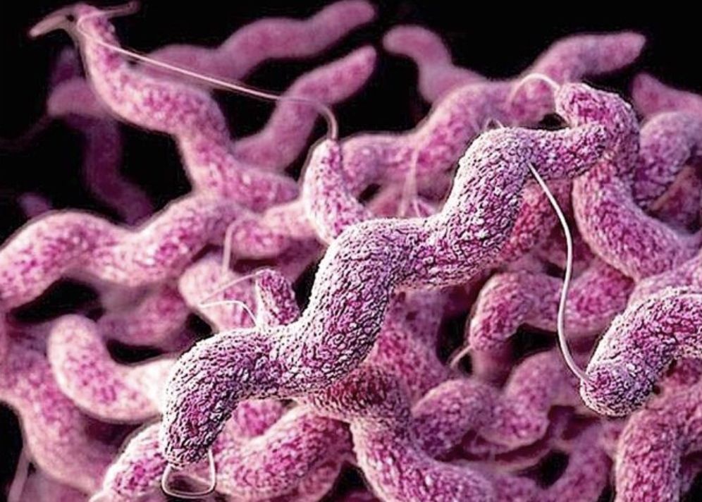
Campylobacter infections


Campylobacter overview


Epidemiology - Most common bacterial cause of gastroenteritis globally (Kaakoush et al., 2015)

  • Primarily Campylobacter jejuni (90% of infections) - Also: C. coli, C. lari, and other species

Characteristics

  • Gram-negative, microaerophilic curved rod

  • Minimal growth on routine culture media

  • Fastidious organism; requires special handling

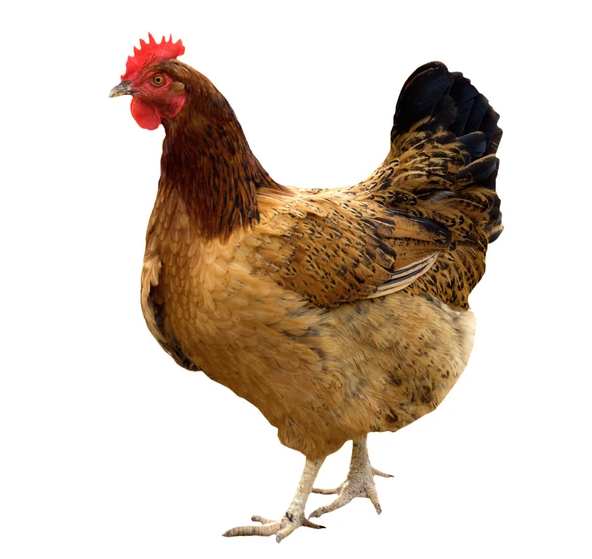
Campylobacter transmission


Animal Reservoir

  • Common commensal in poultry (colonizes GI tract: 50-90% in GI tract)

    • Also found in cattle, pigs, dogs, cats
  • Undercooked meat: primary source - unpasteurized milk: significant source

Environmental Survival

  • Survives in freshwater at temperatures <15°C

  • Sensitive to heat, desiccation, oxygen at room temperature - short survival in food chain; requires careful handling

Direct Transmission

  • Person-to-person transmission: uncommon but documented

  • Fecal-oral route primarily - Animal contact risk factor

Campylobacter clinical features


Incubation Period

  • Mean: 3 days (range 1-7 days) - Longer than many bacterial pathogens

Typical presentation

  • Affects both small and large bowel → mixed diarrhea pattern

  • Watery AND bloody diarrhea common

  • Febrile prodrome in ~1/3 of cases (fever, malaise, myalgias)

  • Abdominal pain often prominent and severe

Systemic complications

  • Bacteremia in 0.1-1% (higher in immunocompromised)

  • Septic arthritis, osteomyelitis, meningitis (rare)

  • Post-infectious syndromes (see next slide)

Campylobacter complications


Post-infectious complications

Guillain-Barré Syndrome (GBS)

  • Estimated 3-40% of GBS cases linked to prior Campylobacter (Nachamkin et al., 1998)

  • Mechanism: molecular mimicry

  • Antibodies cross-react with GM1 ganglioside

  • Ascending paralysis 1-3 weeks after diarrhea

Reactive Arthritis

  • Occurs in 2.6% of infections

  • HLA-B27 association

  • Arthralgia/arthritis weeks after diarrhea

  • Can be prolonged and disabling

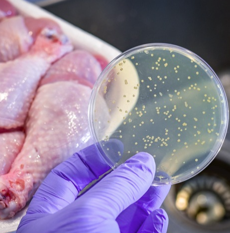
Campylobacter diagnosis


Laboratory Detection

  • Stool culture on selective media (Campy agar, CCDA agar) -

  • Requires microaerophilic conditions

  • Gram-negative, S-shaped or curved rods on microscopy

  • Culture takes 48-72 hours minimum

Molecular Methods

  • PCR increasingly available at reference labs

  • Rapid diagnosis possible

  • Higher sensitivity than culture

Salmonella infections

Salmonella overview


Epidemiology

Diversity

  • >1,400 serotypes identified

  • Two major clinical syndromes: gastroenteritis vs. enteric fever

  • Highest incidence globally: South Asia

Non-typhoidal Salmonella epidemiology


Serotype distribution - S. enteritidis: most common globally

  • S. typhimurium: second most common

  • Both associated with poultry and poultry products

  • Transovarial transmission in hens explains egg contamination

Geographic & seasonal patterns

  • Incidence highest in South and Southeast Asia

  • Seasonal peaks: summer and autumn in temperate climates

  • Year-round in tropical regions

Non-typhoidal Salmonella transmission

Non-typhoidal Salmonella clinical features


Incubation Period : 8-72 hours (typically 12-36 hours)

Typical Presentation :

  • Diarrhea with abdominal pain and cramping

  • Fever in ~50% (often high—>39°C)

  • Nausea and vomiting common

  • Systemic symptoms: malaise, headache

Risk Factors for Severe Disease

  • Extremes of age (<5 or >65 years)
  • Achlorhydria or antacid use, Inflammatory bowel disease
  • Sickle cell disease, immunosuppression

Prognosis

  • Self-limited in immunocompetent hosts, bacteremia in <5% (higher with underlying conditions)

  • Duration typically 4-7 days

Non-typhoidal Salmonella — Asymptomatic Carriage


Chronic Carriers

  • Shedding bacteria >1 year after infection

  • Prevalence: 0.6-2% of infected individuals

  • More common with S. enteritidis than other serotypes

  • Risk factors: female sex, older age, biliary disease

Clinical Implications

  • Potential source for transmission to others - Important for food handlers and healthcare workers

  • Prolonged antibiotics (e.g., fluoroquinolone) may clear carriage

  • Cholecystectomy eradicates infection in some biliary carriers

Enteric/typhoid Fever


Epidemiology : Caused by Salmonella typhi (endemic in South Asia, Africa)

  • Also S. paratyphi (Asia-Pacific region)

  • Humans are the only reservoir - ~21 million cases and 200,000 deaths annually globally

  • Mortality 1-4% with treatment; 20-30% without

Risk Factors for Acquisition - Travel to endemic areas (South Asia especially)

  • Poor sanitation exposure

  • Close contact with chronic carriers

Typhoid fever — Clinical progression

Week 1: Septicemia Phase

  • Gradual fever onset (prodrome over days)
  • High fever develops, continuing to rise
  • Bacteremia present
  • Relative bradycardia (unusual for degree of fever)
  • Malaise, headache, myalgias

Week 2-3: Systemic Phase

  • Sustained high fever (often continuous pattern—“staircase fever”)
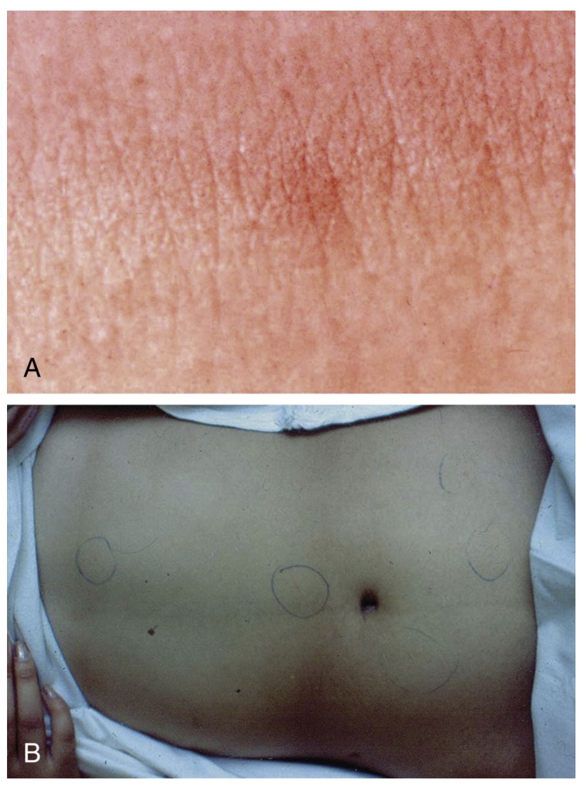
  • Rose spots rash (evanescent, 2-3mm rose-colored papules on trunk)
  • Hepatosplenomegaly with abdominal pain and distension, diarrhea or constipation

Week 3-4: Crisis Phase

  • Risk of intestinal perforation (Peyer’s patches ulcerate)

  • Septic shock possible

  • Delirium and altered mental status (“typhoid state”)

  • Myocarditis, pneumonia

Typhoid fever — Treatment


  • Antimicrobial challenges

    • Fluoroquinolone resistance increasing in South Asia

    • Multidrug-resistant strains (TMP/SMX, chloramphenicol, ampicillin) common

    • Extensively drug-resistant (XDR) strains emerging

  • Treatment options - First-line (susceptible): Fluoroquinolone (ciprofloxacin)

    • Alternatives: Third-generation cephalosporins (ceftriaxone, cefixime)

    • Resistant strains: Azithromycin (5-day course) for nalidixic acid-resistant strains

    • Duration: 7-14 days depending on severity and response

  • Prognosis with treatment - Defervescence typically 4-6 days after starting therapy

    • Relapse possible 1-2 weeks after apparent cure - Follow-up cultures recommended to document clearance

Shigella overview


Microbiology

  • Non-motile gram-negative Enterobacterales

  • Four serogroups: dysenteriae, flexneri, boydii, sonnei (Kotloff et al., 2018)

  • Humans are the only reservoir (crucial difference from Salmonella)

Clinical Significance

  • Third most common bacterial cause of diarrhea (after Salmonella and Campylobacter)

  • Associated with severe dysentery and complications

  • Rapid person-to-person spread in closed environments

Shigella pathophysiology and complications


Virulence Mechanisms

  • Invasion of colonic epithelium (ipaB, ipaC genes)

  • Intracellular multiplication - abscess formation and mucosal ulceration

  • Enterotoxin production: ShET1, ShET2

Complications

  • Shiga toxin produced by S. dysenteriae → HUS possible - HUS occurs in ~8% of children with S. dysenteriae infection

  • Toxic megacolon (rare)

  • Protein-calorie malnutrition from persistent diarrhea

  • Seizures, febrile delirium (especially in children)

Shigella diagnosis and treatment


Laboratory Diagnosis

  • Stool culture on selective media (HE agar, XLD agar)

  • Preferred: culture from mucoid/blood-stained stool

  • Non-motile gram-negative colonies - PCR for Stx gene (S. dysenteriae)

Resistance Patterns

  • Asia/Africa: 20-30% resistance to third-generation cephalosporins

  • TMP/SMX resistance: 65-85% in some regions

  • Fluoroquinolone resistance increasing (esp. S. sonnei in Asia)

Treatment

  • First-line: Fluoroquinolone or ceftriaxone (when fluoroquinolone susceptibility uncertain)

  • Alternative: Azithromycin

  • Duration: 5-7 days

Shigella — When to treat


Standard Recommendation

  • Most infections resolve without antibiotics

  • Treatment doesn’t significantly alter outcomes in mild-moderate disease

Treat When

  • Immunocompromised patients (including HIV)

  • Severe diarrhea or dysentery

  • Bacteremia or extraintestinal infection

  • High risk for transmission (food handlers, daycare workers)

Benefit of Treatment

  • Decreases symptom duration by ~2 days

  • Reduces fecal shedding (may reduce transmission)

  • Prevents complications in vulnerable populations

Yersinia

Species of clinical importance:

  • Yersinia enterocolitica - Yersinia pseudotuberculosis

Key characteristics - Zoonotic infections: wild and domestic animals

  • Transmission: undercooked pork, contaminated water

  • Can survive refrigeration (cold enrichment aids culture)

Clinical features : Watery diarrhea or dysentery

  • Distinctive: pharyngitis in ~20% (pharyngitis-gastroenteritis pattern)

  • Acute mesenteric lymphadenitis: can mimic appendicitis

  • Fever and abdominal pain prominent

  • Can cause arthralgia (particularly HLA-B27 associated)

Lab diagnosis - Culture on selective media (CIN agar)

  • Overgrowth by normal flora; requires selective medium or cold enrichment

John Snow- Birth of medical epidemiology


Vibrio cholerae


Epidemiology

  • Endemic in South Asia (particularly Bangladesh, India) (Sack et al., 2004)
  • Seventh pandemic ongoing since 1961
  • Transmitted via contaminated water in areas with poor sanitation
  • Epidemic potential high; 3-5 million cases, 100,000-300,000 deaths annually (Ali et al., 2015)

Clinical Presentation

  • Acute watery non-bloody diarrhea
  • Characteristic “rice-water stools” (clear, watery, with flecks)
  • Severe dehydration and shock possible
  • Vomiting common
  • Can be fulminant with progression to hypovolemic shock

Cholera management


Diagnostic approach

  • Culture on TCBS (Thiosulfate-Citrate-Bile Salts) agar

  • Oxidase-positive, gram-negative curved rods

  • PCR available at reference labs

Treatment

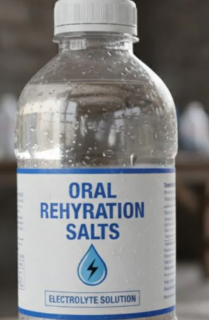
Fluid replacement is paramount!!!

  • Oral rehydration solution (ORS) is first-line

  • IV fluids (normal saline or Ringer’s lactate) for severe dehydration

  • Replacement volumes can be massive (10-20 L/day in severe cases)

  • Monitor for electrolyte abnormalities

Antimicrobial therapy

  • Decreases duration and volume of diarrhea

  • Doxycycline, fluoroquinolones, or azithromycin

  • Secondary to fluid replacement in priority

Prevention - Vaxchora: oral cholera vaccine for travelers to endemic areas

  • Provides ~90% protection for 3 months, wanes thereafter - Food and water precautions essential

Traveler’s Diarrhea

Traveler’s diarrhea — Overview


Epidemiology

  • Affects 300-500 million travelers annually

  • Attack rate varies by destination: 5-50% depending on region (Steffen et al., 2015)

  • Onset typically 5-15 days after arrival in endemic region

  • Duration usually 1-5 days (self-limiting in 90%)

Clinical Presentation

  • Watery diarrhea most common (80%)

  • Some bloody stools possible (10-20%)

  • Fever in 20-30%

  • Cramping abdominal pain

  • Systemic symptoms mild

Definition

  • ≥3 unformed stools in 24 hours plus 1+ GI symptom

  • Occurring in someone traveling to area of higher risk

Traveler’s diarrhea — Etiology by region


Pathogen Frequency Geographic Notes
ETEC 40-50% Most common worldwide
Campylobacter jejuni 5-30% Higher in Asia
Salmonella spp. 5-20% Variable by region
Shigella spp. 5-15% Higher in developing regions
Enteroinvasive E. coli 5-10% Variable
Protozoa (Giardia, Crypto) 2-5% More in rural areas
Viral 5-10% Norovirus, Rotavirus, Adenovirus
Noninfectious 10-20% Dietary changes, altitude

Traveler’s diarrhea — Prevention


Food and water precautions

  • Drink bottled or boiled water

  • Avoid ice, raw vegetables, raw/undercooked meat

  • Peel own fruits

  • Avoid street food and unpasteurized dairy

Antimicrobial prophylaxis

  • Not routinely recommended (resistance, adverse effects)

  • Consider for high-risk patients (immunocompromised, severe underlying disease)

  • Bismuth subsalicylate: effective prophylaxis (2 tablets QID)

  • Duration: maximum 3 weeks

Traveler’s Diarrhea — Self-treatment


Treatment Options

Preferred Approach

  • Azithromycin 500 mg once daily, 3 days (Riddle et al., 2016)

  • Covers ETEC, Campylobacter, Shigella

  • Lower resistance rates than fluoroquinolones

Alternative Approaches

  • Fluoroquinolone (levofloxacin, ciprofloxacin) if available

  • Rifaximin 200 mg TID, 3 days (non-absorbed, minimal resistance)

  • Symptomatic Therapy - Loperamide (Imodium): effective for cramping

    • Combine with antibiotic for faster resolution - Bismuth subsalicylate: both treatment and symptomatic relief

Diarrhea in HIV/AIDS


Epidemiology

  • Affects 30-60% of patients with AIDS (CD4 <200) (Sanchez et al., 2005)

  • Incidence decreased markedly with antiretroviral therapy

  • ART with immune reconstitution reduces diarrheal disease

Infectious Etiologies in AIDS

  • Cryptosporidium parvum: most common parasitic cause

  • Cytomegalovirus: causes ulcerative colitis pattern

  • Microsporidium: can cause chronic diarrhea

  • Mycobacterium avium complex: systemic infection

  • Conventional pathogens remain common (Salmonella, Campylobacter)

Management - Start/optimize antiretroviral therapy (most important) - Ganciclovir for CMV colitis - Multipathogen testing recommended - Empiric therapy based on CD4 count and epidemiology

Diarrhea in transplant recipients


  • Frequency and Timing

    • 50-80% of solid organ transplant (SOT) recipients experience diarrhea

    • Varies by organ type and immunosuppression level -

    • Can occur months to years post-transplant

  • Infectious Etiologies

    • Clostridioides difficile: most common infectious cause (9-20% incidence) (McDonald et al., 2018)

    • Norovirus: prolonged shedding common

    • Cryptosporidium, Giardia, Cyclospora

    • Cytomegalovirus: ulcerative colitis pattern

  • Noninfectious Causes - Up to 66% of cases in some series - medication side effects (mycophenolate, tacrolimus) , sorbitol-containing medications, IBD-like inflammation (idiopathic)

  • Diagnostic Approach - Multipathogen testing essential (stool culture, EIA, PCR)

    • Consider colonoscopy with biopsy - Address immunosuppression optimization

Diarrhea in immunocompromised patients — General approach


  • Key Diagnostic Principles

    • Identify etiologic agent whenever possible (broad differential)

    • Multipathogen testing: stool culture, parasitic studies, molecular panel

    • Lower threshold for colonoscopy and biopsy

    • Consider unusual pathogens based on immune defect

  • Treatment Considerations

    • Pathogen-specific therapy when identified

    • Avoid empiric broad-spectrum antibiotics when possible

    • Address underlying immune defect (ART, immunosuppression optimization)

    • Monitor for immune recovery inflammation (MAC disease, IRIS)

Hospital-acquired diarrhea


  • Epidemiology: Occurs in 10-15% of hospitalized patients

    • Clostridioides difficile: most common infectious cause (Lessa et al., 2015)

    • Associated with increased morbidity, mortality, and healthcare costs

  • Risk factors: Recent or current antimicrobial therapy (strongest risk factor)

    • Advanced age

    • Severity of underlying illness

    • Prolonged hospitalization

    • Immunosuppression

  • Clinical features : Occurs after ≥3 days hospitalization

    • Watery diarrhea most common

    • Fever, leukocytosis, abdominal pain -

    • Can progress to toxic megacolon or perforation

  • C. difficile-associated disease

    • Toxin-mediated disease (toxin A, toxin B) -Antimicrobial exposure disrupts normal flora

    • Transmission via spores: contact precautions required (hand sanitizer not sufficient)

    • Increasing incidence of severe, recurrent disease

Diarrhea in institutional settings


  • Long-Term Care Facilities

    • One-third of residents experience diarrhea annually

    • C. difficile most common

    • Rotavirus, G. lamblia seasonal outbreaks

    • Norovirus rapid spread in winter

    • Nutritional impact: worsens outcomes in elderly

  • Daycare and School Settings

    • Rotavirus common in young children (prior to universal vaccination)

    • G. lamblia outbreaks in daycare

    • Shigella spread via fecal-oral route

    • ETEC in contaminated water/food

    • Exclusion policies important for control

  • Neonatal diarrhea

    • Often caused by EPEC serotypes

    • Risk of severe dehydration in newborns

    • Historical mortality 24-50%; now <5% with rehydration therapy

    • Insidious onset; requires high clinical suspicion - May present with failure to thrive

Principles of treatment — Rehydration


  • Oral Rehydration Solution (ORS)

    • First-line for mild-moderate dehydration

    • WHO-recommended formulation: sodium 75 mmol/L, glucose 75 mmol/L, chloride 65 mmol/L, potassium 20 mmol/L

    • Effective for >90% of acute diarrhea cases

  • Rehydration Approach

    • Replace ongoing losses (10 mL/kg per stool)

    • Add maintenance fluids

    • Early rehydration prevents severe dehydration

    • Resume age-appropriate diet early

  • IV Rehydration - Reserved for severe dehydration, vomiting, shock

    • Normal saline or Ringer’s lactate preferred

    • Careful electrolyte monitoring - Transition to oral when feasible

Symptomatic treatment


  • Bismuth Subsalicylate

    • Reduces diarrheal volume by 30-50%

    • Antimicrobial properties against several pathogens

    • Useful for both prophylaxis and treatment

    • Avoid in salicylate allergy; concern for drug interactions - Useful in traveler’s diarrhea management

  • Antimotility Agents: Use with Caution - Loperamide (Imodium): effective for cramping

    • Risk: can precipitate toxic megacolon (contraindicated in bloody diarrhea, fever, severe disease)

    • Never use with suspected EHEC

    • Combined with antibiotics: effective for traveler’s diarrhea

    • Generally safe in mild, watery, non-inflammatory diarrhea

  • Probiotics

    • Insufficient evidence for general recommendation

    • May be role in specific contexts (antibiotic-associated diarrhea)

    • Not harmful but not proven beneficial in most diarrhea

When to use antibiotics

  • Empiric Antibiotics Recommended For:

    • Severe diarrhea (bloody, fever, >8 stools/day)

    • Diarrhea in immunocompromised patients

    • Traveler’s diarrhea (if symptomatic treatment not effective)

    • Suspected invasive pathogen (Salmonella bacteremia, Shigella in systemically ill)

    • Institutional outbreaks (control transmission)

  • Avoid Antibiotics: Suspected or confirmed EHEC/STEC (increases HUS risk)

    • Viral diarrhea - Mild, watery, non-bloody diarrhea in immunocompetent hosts

    • Non-typhoidal Salmonella gastroenteritis in most patients

First-Line Empiric Choices

  • Azithromycin 500 mg daily × 3 days (preferred for traveler’s diarrhea)

  • Fluoroquinolone (ciprofloxacin 500 mg BID × 3 days) where resistance is low

  • Adjust based on local resistance patterns

Micronutrient supplementation


  • Zinc Supplementation

    • Particularly in children <5 years in developing countries (Lazzerini and Wanzira, 2016)

    • Reduces duration and severity of diarrhea

    • Decreases risk of subsequent infections for 2-3 months

    • Dose: 10-20 mg elemental zinc daily for 10-14 days

    • Strong evidence supports benefit, especially in malnourished children

  • Other Micronutrients

    • Vitamin A: benefit in deficient populations

    • Iron: avoid during acute infection (may worsen)

    • Folate, B vitamins: supportive during recovery

Role in Developed Countries - Less emphasis (better nutritional status baseline) - Consider in malnourished or vulnerable populations - Not harmful if administered

Key Take-Home Messages

  1. Most diarrhea is viral and self-limited — reserve specific testing and antibiotics for cases suggesting bacterial infection

  2. Rehydration is the cornerstone of treatment — ORS is highly effective and first-line for most cases (Munos et al., 2010)

  3. Identify patients needing hospitalization or antibiotics — use clinical features, history, and exam to guide severity assessment and testing

  4. EHEC/STEC demands special attention — antibiotics are contraindicated and increase HUS risk

  5. Geographic and risk-factor epidemiology matters — tailor diagnostic approach and empiric therapy based on exposure history and patient factors

Clinical decision Framework

① Assess Severity
Dehydration  •  Vital signs  •  Systemic symptoms
⚠ Red flags: bloody stools  |  fever >39°C  |  altered mental status  |  shock
② Targeted History
Travel  •  Food/water exposure  •  Sick contacts  •  Immunocompromise  •  Recent antibiotics  •  Medications
③ Decide on Testing
✓ Send Testing
• Bloody diarrhea
• Fever >38.5°C
• Systemic illness
• Immunocompromised
• Duration >7 days
✗ No Testing Needed
• Mild watery diarrhea <7 days
• No red flags
• Immunocompetent host
④ Treatment
Rehydration — first-line for all     Antibiotics — only if bacterial criteria met; avoid for EHEC     Symptomatics — if no contraindications

References

  • Troeger C, et al. Estimates of the global, regional, and national morbidity, mortality, and aetiologies of diarrhea in 195 countries: a systematic analysis for the Global Burden of Disease Study 2016. Lancet Infect Dis. 2018.

  • Liu L, et al. Global, regional, and national causes of child mortality in 2000-13, with projections to inform post-2015 priorities. Lancet. 2015.

  • Freedman SB, et al. Pediatric Gastroenteritis in Developed and Developing Countries. Gastroenterology. 2020.

  • Platts-Mills JA, et al. Pathogen-specific burdens of community diarrhoea in developing countries. Lancet Glob Health. 2015.

Ahmed SM, Hall AJ, Robinson AE, et al. Global prevalence of norovirus in cases of gastroenteritis: A systematic review and meta-analysis. The Lancet Infectious Diseases 2014;14:725–30. https://doi.org/10.1016/S1473-3099(14)70767-4.
Ali M, Nelson AR, Lopez AL, Sack DA. Updated global burden of cholera in endemic countries. PLoS Neglected Tropical Diseases 2015;9:e0003832. https://doi.org/10.1371/journal.pntd.0003832.
Crump JA, Mintz ED. Global trends in typhoid and paratyphoid fever. Clinical Infectious Diseases 2010;50:241–6. https://doi.org/10.1086/649541.
Kaakoush NO, Castaño-Rodríguez N, Mitchell HM, Man SM. Global epidemiology of Campylobacter infection. Clinical Microbiology Reviews 2015;28:687–720. https://doi.org/10.1128/CMR.00006-15.
Karch H, Tarr PI, Bielaszewska M. Enterohaemorrhagic Escherichia coli in human medicine. International Journal of Medical Microbiology 2005;295:405–18. https://doi.org/10.1016/j.ijmm.2005.06.009.
Kotloff KL, Riddle MS, Platts-Mills JA, et al. Shigellosis. The Lancet 2018;391:801–12. https://doi.org/10.1016/S0140-6736(17)33296-8.
Lazzerini M, Wanzira H. Oral zinc for treating diarrhoea in children. Cochrane Database of Systematic Reviews 2016;12:CD005436. https://doi.org/10.1002/14651858.CD005436.pub5.
Lessa FC, Mu Y, Bamberg WM, et al. Burden of Clostridium difficile infection in the United States. New England Journal of Medicine 2015;372:825–34. https://doi.org/10.1056/NEJMoa1408913.
Liu J, Platts-Mills JA, Juma J, et al. Use of quantitative molecular diagnostic methods to identify causes of diarrhoea in children: A reanalysis of the GEMS case-control study. The Lancet 2016;388:129–301. https://doi.org/10.1016/S0140-6736(16)31529-X.
Majowicz SE, Musto J, Scallan E, et al. The global burden of nontyphoidal Salmonella gastroenteritis. Clinical Infectious Diseases 2010;50:882–9. https://doi.org/10.1086/650733.
McDonald LC, Gerding DN, Johnson S, et al. Clinical practice guidelines for Clostridium difficile infection in adults and children: 2017 update by the IDSA and SHEA. Clinical Infectious Diseases 2018;66:e1–48. https://doi.org/10.1093/cid/cix1085.
Munos MK, Walker CLF, Black RE. The effect of oral rehydration solution and recommended home fluids on diarrhoea mortality. International Journal of Epidemiology 2010;39:i75–87. https://doi.org/10.1093/ije/dyq025.
Nachamkin I, Allos BM, Ho T. Campylobacter species and Guillain-Barré syndrome. Clinical Microbiology Reviews 1998;11:555–67. https://doi.org/10.1128/CMR.11.3.555.
Parashar UD, Gibson CJ, Bresee JS, Glass RI. Rotavirus and severe childhood diarrhea. Emerging Infectious Diseases 2006;12:304–6. https://doi.org/10.3201/eid1202.050006.
Patel MM, Widdowson MA, Glass RI, et al. Systematic literature review of role of noroviruses in sporadic gastroenteritis. Emerging Infectious Diseases 2008;14:1224–31. https://doi.org/10.3201/eid1408.071114.
Qadri F, Svennerholm AM, Faruque ASG, Sack RB. Enterotoxigenic Escherichia coli in developing countries: Epidemiology, microbiology, clinical features, treatment, and prevention. Clinical Microbiology Reviews 2005;18:465–83. https://doi.org/10.1128/CMR.18.3.465-483.2005.
Rasko DA, Webster DR, Sahl JW, et al. Origins of the E. coli strain causing an outbreak of hemolytic-uremic syndrome in Germany. New England Journal of Medicine 2011;365:709–17. https://doi.org/10.1056/NEJMoa1106920.
Riddle MS, Connor BA, Beeching NJ, et al. ACG clinical guideline: Diagnosis, treatment, and prevention of acute diarrheal infections in adults. American Journal of Gastroenterology 2016;111:602–22. https://doi.org/10.1038/ajg.2016.126.
Ruiz-Palacios GM, Pérez-Schael I, Velázquez FR, et al. Safety and efficacy of an attenuated vaccine against severe rotavirus gastroenteritis. New England Journal of Medicine 2006;354:11–22. https://doi.org/10.1056/NEJMoa052434.
Sack DA, Sack RB, Nair GB, Siddique AK. Cholera. The Lancet 2004;363:223–33. https://doi.org/10.1016/S0140-6736(03)15328-7.
Sanchez TH, Brooks JT, Sullivan PS, et al. Bacterial diarrhea in persons with HIV infection, United States, 1992–2002. Clinical Infectious Diseases 2005;41:1621–7. https://doi.org/10.1086/498027.
Steffen R, Hill DR, DuPont HL. Traveler’s diarrhea: A clinical review. JAMA 2015;313:71–80. https://doi.org/10.1001/jama.2014.17373.
Tarr PI, Gordon CA, Chandler WL. Shiga-toxin-producing Escherichia coli and haemolytic uraemic syndrome. The Lancet 2005;365:1073–86. https://doi.org/10.1016/S0140-6736(05)71144-2.
Tate JE, Burton AH, Boschi-Pinto C, Parashar UD. WHO-coordinated Global Rotavirus Surveillance Network. Global, regional, and national estimates of rotavirus mortality in children <5 years of age, 2000–2013. Clinical Infectious Diseases 2016;62:S96–105. https://doi.org/10.1093/cid/civ1013.
Troeger C, Blacker BF, Khalil IA, et al. GBD 2016 Diarrhoeal Diseases Collaborators. Estimates of the global, regional, and national morbidity, mortality, and aetiologies of diarrhoea in 195 countries: A systematic analysis for the Global Burden of Disease Study 2016. The Lancet Infectious Diseases 2018;18:1211–28. https://doi.org/10.1016/S1473-3099(18)30362-1.
Vesikari T, Matson DO, Dennehy P, et al. Safety and efficacy of a pentavalent human-bovine (WC3) reassortant rotavirus vaccine. New England Journal of Medicine 2006;354:23–33. https://doi.org/10.1056/NEJMoa052664.