Fever of Unknown Origin

Russell E. Lewis

2026-03-01

Fever of Unknown Origin (FUO)


Prof. Russell E. Lewis
Department of Molecular Medicine
University of Padua


russelledward.lewis@unipd.it
https://github.com/Russlewisbo


slides available at: www.padovaid.com

Objectives


  • Recognize leading infectious causes of FUO in key patient groups
  • Identify fever patterns and clinical histories that may direct diagnosis
  • Differentiate FUO risks and possible spectrum of pathogens in immunocompromised hosts

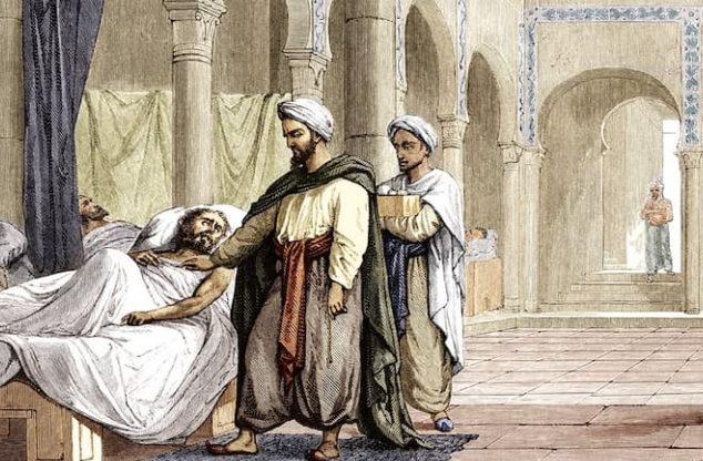
The history of fever


  • 10th Century BCE Persian Physician Akhawayni defined a system for fever curves in Hidāyat al-Muta’allimīn fī al-Tibb (The Student’s Handbook of Medicine)
  • Hippocratic physicians proposed that body temperature, and physiologic harmony in general, involved a delicate balance among four corporal humors—blood, phlegm, black bile, and yellow bile.
    • Fever was due to excess of yellow bile (many infections caused jaundice)
  • Galen: many types of fever developed from putrefaction of humors.
  • Middle ages: demonic possession
  • 18th century (Harvey’s discovery of circulation)- friction of blood flow through body causing fermentation and putrefaction in intestines
  • Claude Bernard in the 19th century- metabolic processes in the body


Febris - Roman Goddess of Fever

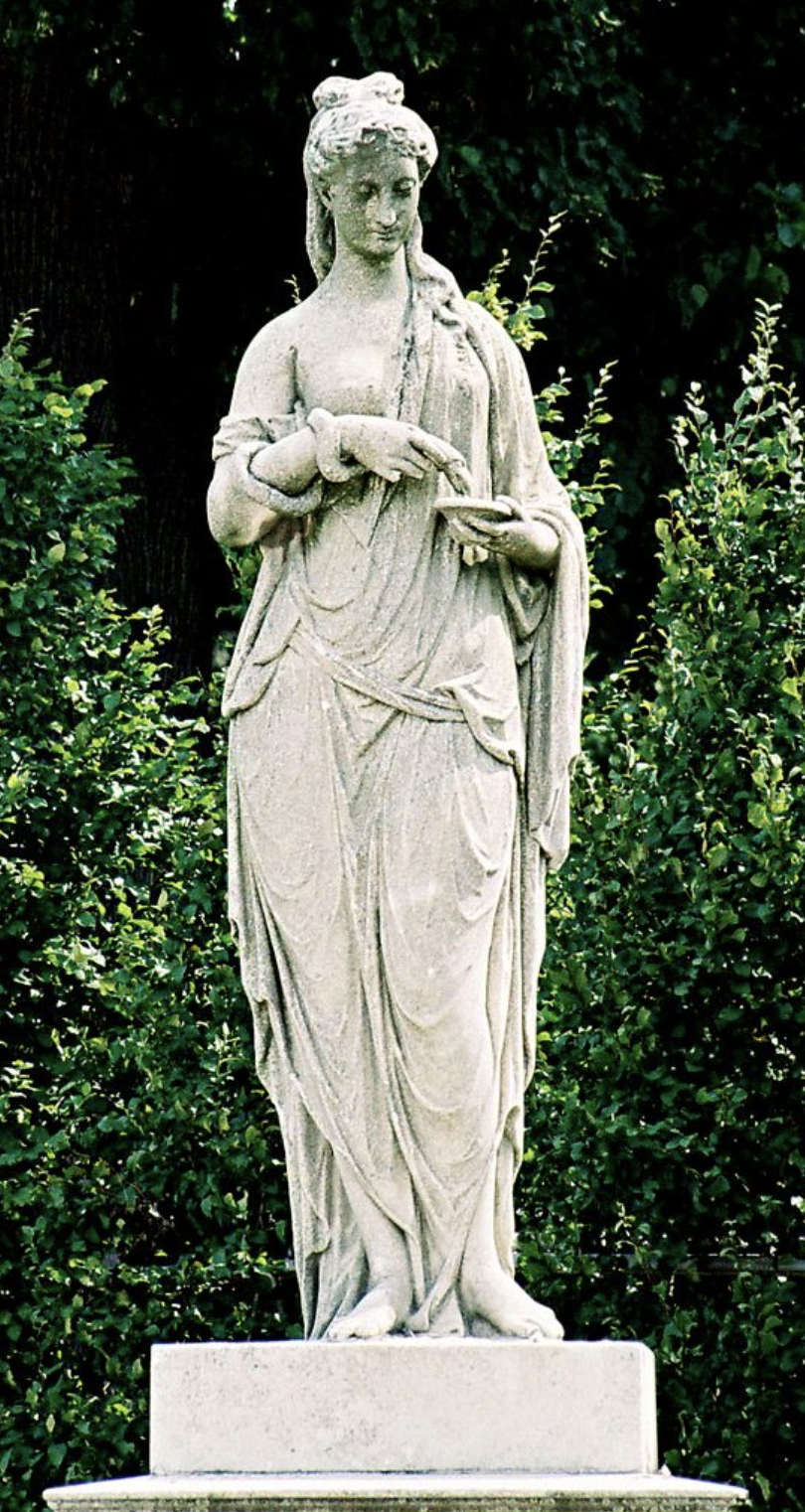
The legend of Febris was said to center around the haunting marshes of Camagna in Southern Italy where like clockwork every year, the people would become deathly ill with a mysterious disease. She was so feared by the Romans that the suffering population had created a cult to Febris. They went so far as to wear protective amulets and build her temples in order to worship her to win her favour.

Early thermometers

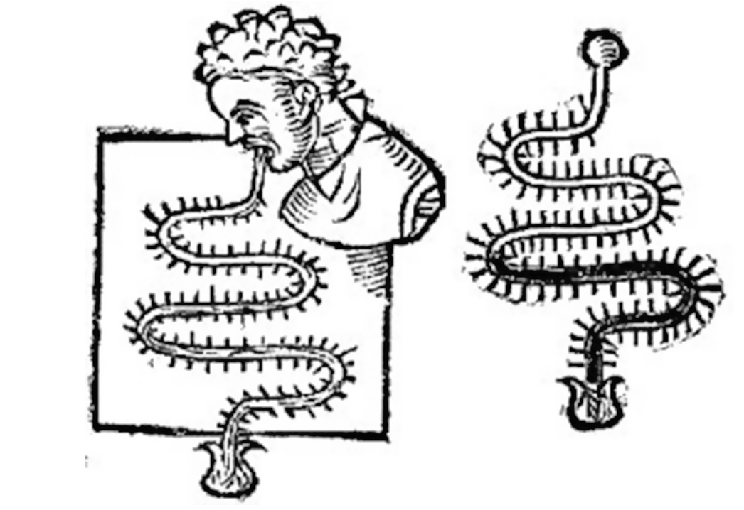
A typical design of a thermoscope is a tube in which a liquid rises and falls as the temperature changes. The Sanctorius thermoscope. Source: Professor Francis Ring, the University of Leeds

Fever in modern medicine


  • Wunderlich’s pioneering studies of thermometry reported normal temperature at 37°C

  • Since the 19th century, humans have become gradually colder - 0.05° to 0.5°C per decade!

    • Current normal range is 36.3 to 36.5°C
    • Less manual labor, less chronic inflammation-infection, poor dental health, increased body mass
  • Mackowiak (1992): mean oral temperature 36.8 ± 0.4°C; only 8% had 37°C

  • Fever now defined as: early-morning temperature ≥37.2°C or anytime ≥ 37.8°C
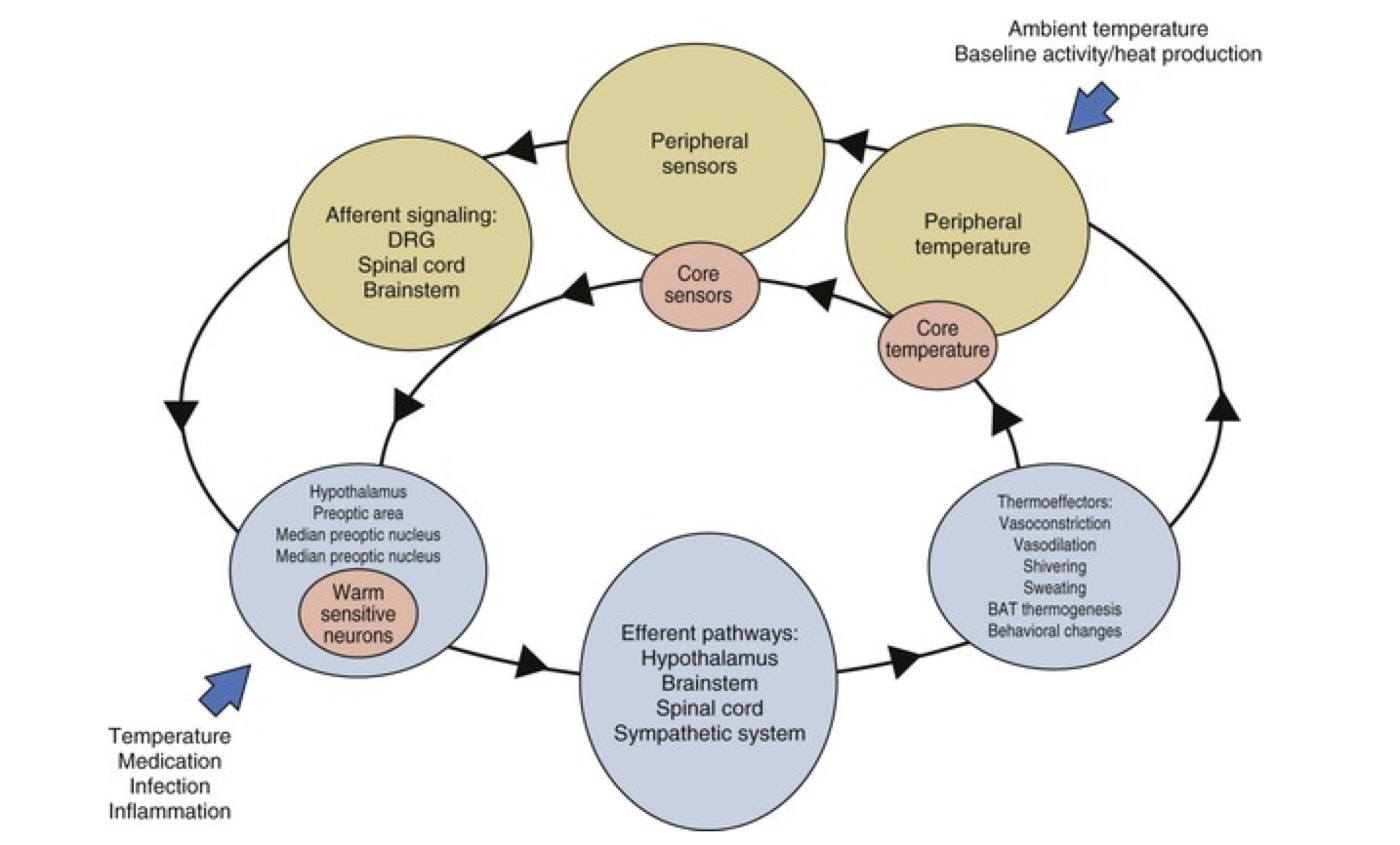
Carl Reinhold Wunderlich

Carl Reinhold Wunderlich

Thermal homeostasis

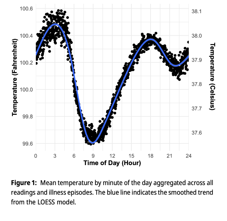
Diurinal pattern of body temperature

Physiologic variables affecting body temperature


  • Age and sex differences
    • Women ~0.5°C higher at ovulation
    • Infants have higher baseline temperatures
  • Circadian rhythm
    • Zenith: late afternoon (peak)
    • Nadir: early morning (trough)
  • Other factors
    • Exercise and physical exertion
    • Medications (antipyretics, corticosteroids)
    • Digestion and recent meals
    • Chronic disease and metabolic conditions

Fever vs. Hyperthermia


Feature Fever Hyperthermia
Set point Elevated Normal
Mechanism Regulated response Unregulated heat generation
Cause Pyrogens (LPS, IL-1, TNF) Heat exposure, drugs, malignant hyperthermia
Pathophysiology Altered hypothalamic setpoint Failure of heat dissipation
Sweating Absent initially (shivering);
later excessive
May be absent
Treatment response Antipyretics effective Antipyretics ineffective; cooling required
Examples Infection, NIID, malignancy Heat stroke, neuroleptic malignant syndrome

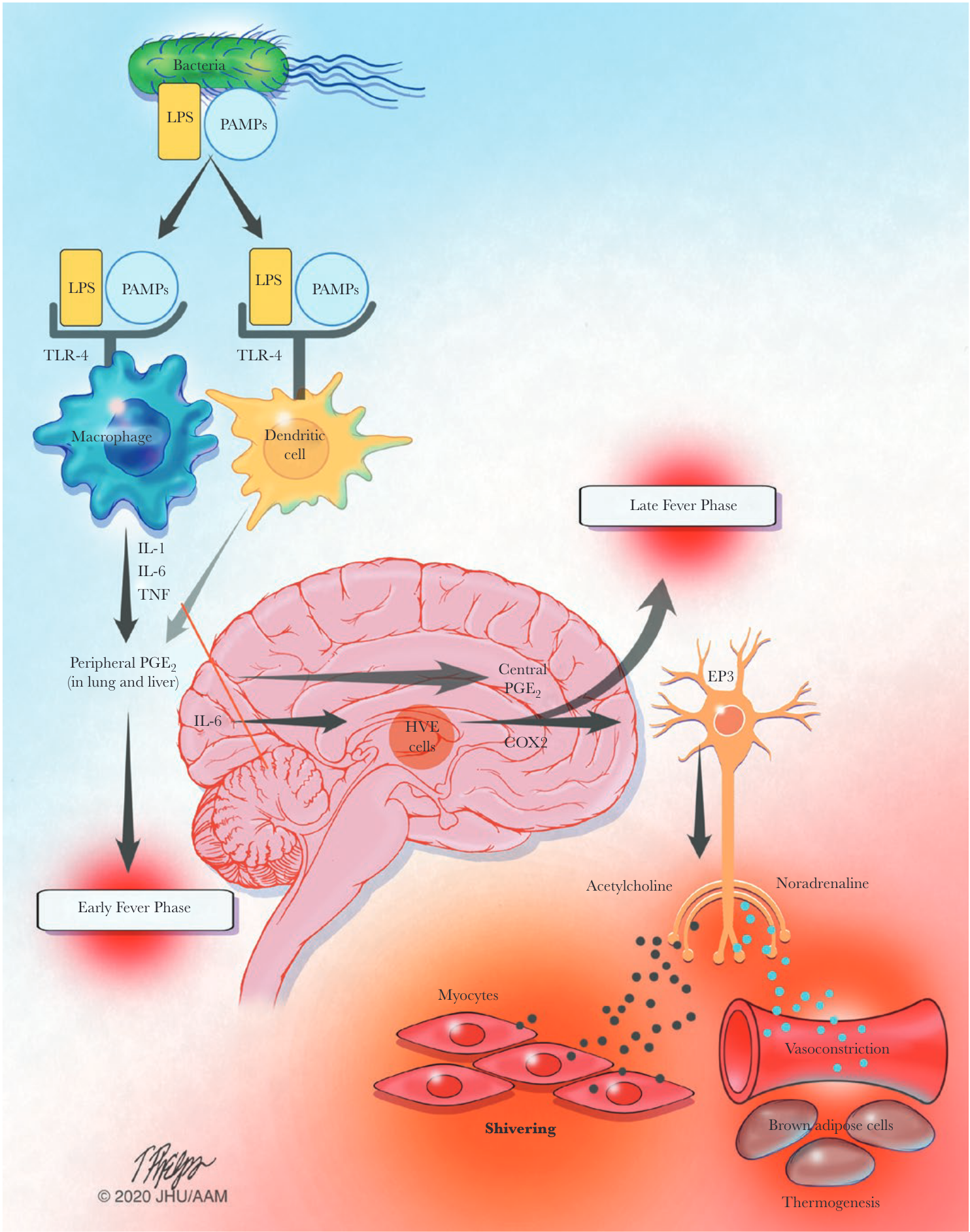
Infection-associated fever - The febrile response


Sequelae of fever - Benefits of fever


  • Phylogenetic conservation suggests fever is beneficial - appears in ectotherms and endotherms
  • Microbial metabolism - most pathogenic bacteria are mesophiles (optimal growth at 35-37°C)
    • Even modest temperature elevations slow replication
  • Iron sequestration - fever triggers hepatic synthesis of hepcidin and lactoferrin
    • Sequesters free iron necessary for bacterial replication
  • Enhanced immune function
    • Increased neutrophil migration
    • Augmented antibody production
    • Enhanced T-cell proliferation

Acute phase proteins


Category Proteins
↑ Complement system C3, C4, C9, Factor B, C1 inhibitor, C4b-binding protein, Mannose-binding lectin
↑ Coagulation / fibrinolysis Fibrinogen, Plasminogen, tPA, Urokinase, Protein S, Vitronectin, PAI-1
↑ Antiproteases α₁-Protease inhibitor, α₁-Antichymotrypsin, Pancreatic trypsin inhibitor, Inter-α-trypsin inhibitors
↑ Transport proteins Ceruloplasmin, Haptoglobin, Hemopexin
↑ Inflammatory mediators Secreted PLA₂, LPS-binding protein, IL-1 receptor antagonist, G-CSF
↑ Others CRP, Serum amyloid A, α₁-Acid glycoprotein, Fibronectin, Ferritin, Angiotensinogen
↓ Negative acute-phase Albumin, Transferrin, Transthyretin, α₂-HS glycoprotein, Alpha-fetoprotein, Thyroxine-binding globulin, IGF-I, Factor XII

Acute phase phenomena


Category Phenomena
Neuroendocrine Fever, somnolence, anorexia; ↑ CRH, corticotropin & cortisol; ↑ arginine vasopressin; ↓ IGF-I; ↑ adrenal catecholamines
Hematopoietic Anemia of chronic disease; leukocytosis; thrombocytosis
Metabolic Muscle loss & negative nitrogen balance; ↓ gluconeogenesis; osteoporosis; ↑ hepatic lipogenesis; ↑ adipose lipolysis; ↓ lipoprotein lipase activity; cachexia
Hepatic ↑ Metallothionein, iNOS, heme oxygenase, MnSOD, TIMP-1; ↓ phosphoenolpyruvate carboxykinase activity
Nonprotein plasma Hypozincemia, hypoferremia, hypercupremia; ↑ plasma retinol & glutathione

Epidemiology of FUO - Definitions

Category Key causes / definition
Classic FUO Infection (TB, endocarditis, occult abscess, zoonoses, enteric fever, syphilis, histoplasmosis), malignancy, autoimmune/autoinflammatory, miscellaneous; includes virally suppressed HIV (CD4 >200)
Nosocomial FUO FUO arising in hospitalized patients
 ICU Bacteremia, pneumonia, C. difficile, fungemia, catheter infections, PE, acalculous cholecystitis, drug fever, stroke/intracranial hemorrhage
 Non-ICU Similar to ICU causes; patient not critically ill
Immunodeficiency-associated FUO Highly variable; depends on type and degree of immunodeficiency
 Organ-transplant recipients Viruses, donor-derived infections, Strongyloides hyperinfection, opportunistic fungi, rejection, GVHD, HLH, ureaplasma hyperammonemia
 Neutropenia Febrile >5 days despite empirical antibiotics; influenced by neutropenia duration, GVHD prophylaxis, antimicrobial agents
 HCT recipients Pre-engraftment: neutropenic causes; early post-engraftment: herpesvirus, adenovirus, hyperacute GVHD, pneumonia; late: relapsed cancer, immune reconstitution
 HIV/AIDS (no ART) Acute retroviral syndrome, mycobacteria, endemic mycoses, toxoplasmosis, cryptococcosis, HHV-8–associated diseases, lymphoma
Travel-associated FUO Malaria, enteric fever, leptospirosis, viral hemorrhagic fevers, typhus, undifferentiated tropical febrile illness

Fever therapy to treat neurosyphilis


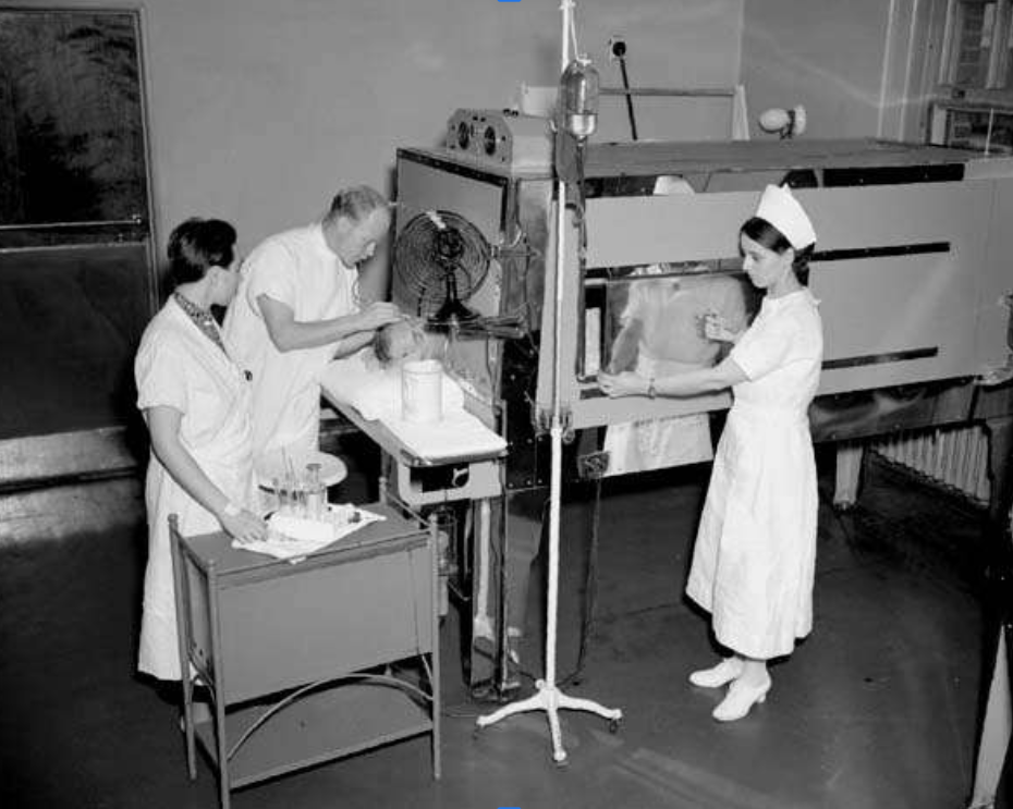
  • Treponema pallidum is uniquely sensitive to increased temperatures

  • Fever therapy involved purposely infecting the patient with Plasmodium vivax with control of the infection with anti- malarians while maintaining the fever it causes to the detriment of other, ongoing, and then-incurable infections such as late-stage syphilis

  • This type of pyrotherapy was most famously used by psychiatrist Julius Wagner-Jauregg, who won the Nobel Prize for Medicine in 1927 for his elaboration of the procedure in treating neurosyphilis

Classic FUO


  • Definition:
    • Temperature of > 38.3°C > 3 weeks
    • Fever >2 separate outpatient visits with diagnostic investigations or
    • Fever >2 visits in hospital of 3 days with diagnostic investigations
      • However,… these definitions are largely subjective
  • Leading causes:
    • Infections (geography dependent)
    • Inflammatory conditions (age dependent)
    • Cancer (age dependent)
    • Undiagnosed/unknown

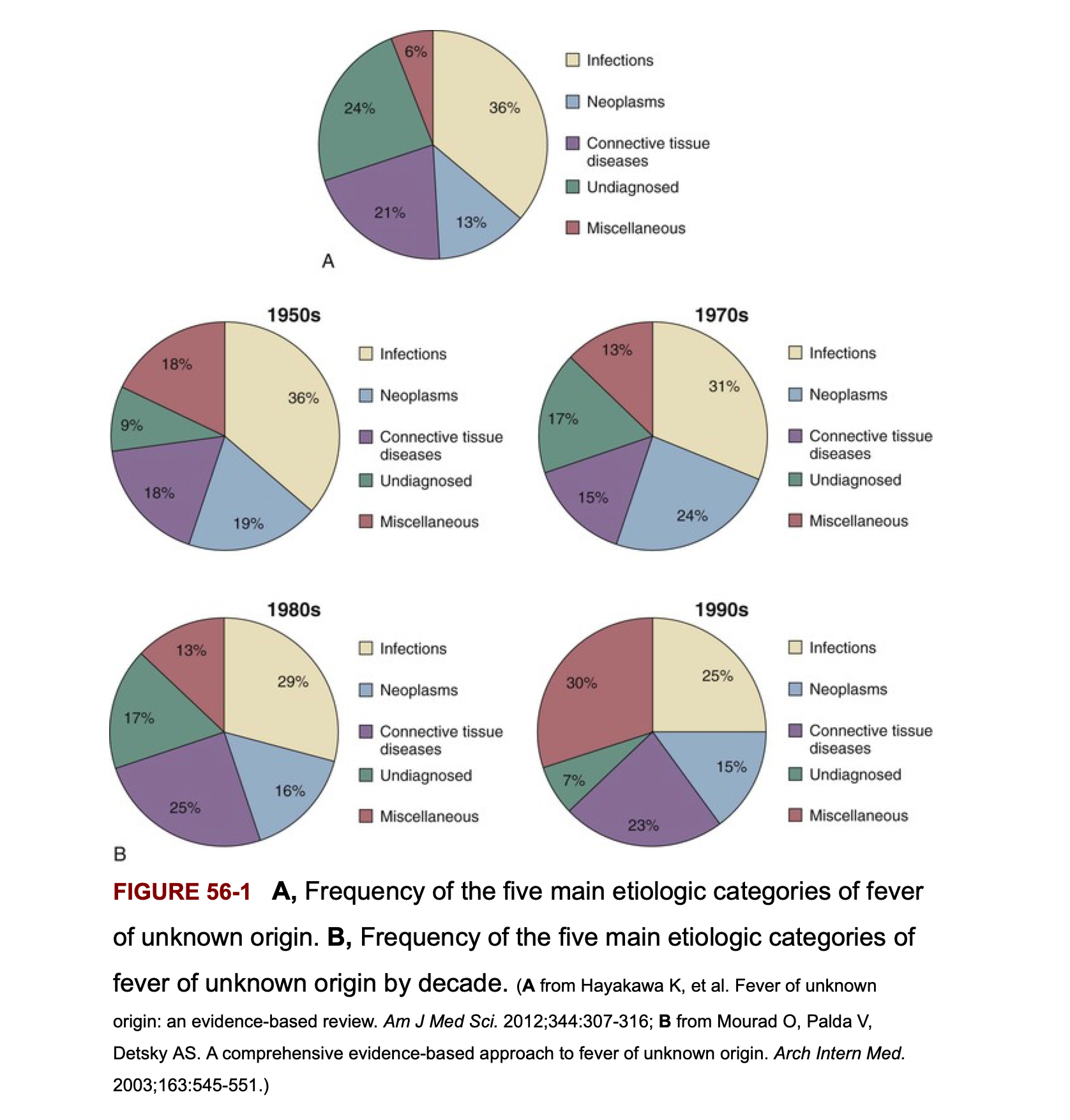
Frequency of the 5 main etiologic categories of FUO

Infectious causes decrease in patients above age 65 years

Classic FUO - Infectious Etiology


  • Chronic or relapsing infections
    • Occult abscess (intra-abdominal, hepatic, splenic, renal, perirectal)
    • Endocarditis (both native and prosthetic valve)
    • Tuberculosis (pulmonary and extrapulmonary)
    • Complicated urinary tract infections (pyelonephritis, prostatitis)
    • Osteomyelitis (especially vertebral)
    • Other: brucellosis, leptospirosis, syphilis, bartonellosis

Rare and miscellaneous causes of fever


Category Examples
Autoinflammatory / periodic fevers Adult-onset Still’s disease, Behçet’s syndrome, Familial Mediterranean fever, Familial Hibernian fever, Periodic fever, Schnitzler’s syndrome
Vascular / cardiac Aortic dissection, Aortitis, Atrial myxoma, Giant coronary aneurysm, Pericarditis, Postpericardiotomy syndrome, Polyarteritis nodosa, Pulmonary emboli, Veno-occlusive disease
Hematologic / oncologic Castleman’s disease, Cyclic neutropenia, Hemoglobinopathies, Hemolytic anemias, Hemophagocytic syndrome, Histiocytosis X, Immunoblastic lymphadenopathy, Lymphomatoid granulomatosis, Myeloproliferative syndromes, Paroxysmal hemoglobinurias, Rosai-Dorfman disease, TTP
Granulomatous / lymphoproliferative Allergic alveolitis, Granulomatous hepatitis, Granulomatous peritonitis, Kikuchi-Fujimoto disease, Lofgren syndrome, Retroperitoneal fibrosis, Sarcoidosis, Subacute necrotizing lymphadenitis
Autoimmune / rheumatologic Autoimmune cholangitis, Erythema multiforme, Inflammatory bowel disease, Serum sickness, Sjögren’s syndrome, Wegener’s granulomatosis
Endocrine / metabolic Addison’s disease, Fabry’s disease, Parathyroid apoplexy, Pheochromocytoma, Thyroiditis & thyrotoxicosis, Vitamin B₁₂ deficiency
GI / hepatic Alcoholic hepatitis, Cirrhotic fever, Pancreatitis
Infectious Bartonellosis, Carcinomatous meningitis, Chronic meningitis, Hantavirus, Human picornavirus, Hypereosinophilic syndrome, Infected urachal cyst, Sinusitis, Whipple’s disease
Other Drug fever & hypersensitivities, Factitious fever, Metal fume fever, Resorbing hematoma

Uncommon and rare causes of FUO


Category Examples
Infections Bartonellosis, hantavirus, coccidioidomycosis, histoplasmosis, blastomycosis, cysticercosis
NIID Addison disease, Behçet syndrome, SLE, vasculitis, inflammatory bowel disease, GPA, sarcoidosis, Sjögren syndrome, thyroiditis, adult-onset Still disease
Malignancy Atrial myxoma, lymphoma, leukemia, pheochromocytoma, Schnitzler syndrome
Other Cirrhotic fever, drug fever, factitious fever, vitamin B₁₂ deficiency, pulmonary embolism

Classic FUO in infants and children


  • Respiratory tract infections - viral, atypical organisms
  • Other infections:
    • UTIs (especially important in young girls)
    • Brucellosis, tuberculosis, bartonellosis
  • Systemic inflammatory conditions:
    • Kawasaki disease (critical, age < 5 years)
    • Inflammatory bowel diseases
    • Still’s disease (juvenile rheumatoid arthritis)
  • Note: Connective tissue diseases and cancers are generally rare in children
  • Important: Joint involvement is a sign of serious disorder - consider endocarditis, leukemia, connective tissue disease

Classic FUO in elderly patients


  • In developed countries: connective tissue diseases > infections
    • Temporal arteritis (critical diagnosis - risk of blindness)
    • Polymyalgia rheumatica
    • Other vasculitides
  • Diagnostic challenge: symptoms are subacute and non-specific, easily attributed to aging
  • Infections still need to be considered:
    • Intra-abdominal abscess
    • Complicated UTIs (often without pyuria)
    • Tuberculosis (may be reactivation)
    • Endocarditis (prosthetic valves more common in elderly)
  • Malignancy: solid tumors and hematologic malignancies increase with age

Fever in returning travelers


Diagnosis Maclean et al (n = 587) Doherty et al (n = 195)
Malaria 32% 42%
Respiratory tract infection 11% 2.6%
Dysentery 4.5% 5.1%
Urinary tract infection / pyelonephritis 4% 2.6%
Dengue fever 2% 6.2%
Enteric fever 2% 1.5%
Hepatitis 6% 3%
Tuberculosis 1% 2%
Rickettsial infection 1% 0.5%
Amebic liver abscess 1% 0%
Acute HIV infection 0.3% 1%
Other miscellaneous infections 4.3% 9.2%
Miscellaneous noninfectious causes 6% 1%
Undiagnosed 25% 24.6%

Nosocomial (Health-Care Associated) FUO


  • Leading causes:
    • Drug fever (especially to antibiotics, anti-epileptic medications)
    • Post-operative complications (e.g., occult abscess, anastomotic leak)
    • Decubitus ulcers with superimposed infection
    • Septic thrombophlebitis (peripherally inserted central catheters)
    • Recurrent pulmonary emboli
    • Myocardial infarction
    • Hematologic malignancy (newly diagnosed during hospitalization)
    • Blood transfusion reaction
    • Reactions to contrast media used in radiologic procedures
    • Clostridium difficile colitis

Fever in post-operative patients


  • Epidemiology: > 1/3 of patients manifest fever in first 5 days post-surgery
  • Infectious vs. non-infectious: < 10% of febrile patients have an identified source or positive cultures
  • Pathophysiology: Fever may represent a physiological response to surgically-induced tissue injury
    • Release of pyrogenic cytokines and interleukins
    • Not necessarily indicative of infection
  • Clinical pearl: Early fever (post-op day 1-3) is usually non-infectious; later fever (day 5+) warrants infection investigation
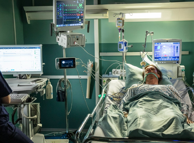
FUO in ICU patients


  • Early fevers are common and often non-infectious
    • Associated with good prognosis
    • Related to inflammatory response to critical illness
  • Prolonged fever carries a poorer prognosis
  • Common complications:
    • Sinusitis (from mechanical ventilation, supine positioning, feeding tubes)
    • Ventilator-associated pneumonia
    • Catheter-related bloodstream infections
  • Other causes similar to nosocomial FUO
    • Abscess, drug fever, septic thrombophlebitis, pulmonary emboli


FUO in stroke patients


  • Non-infective fevers are common in stroke patients
    • Occur earlier after stroke than infection
    • Related to hypothalamic injury and cytokine release
  • Infection-related complications:
    • UTI (from urinary catheterization)
    • Aspiration pneumonia
    • DVT/PE
  • Clinical challenge: Distinguishing fever from infarct-related inflammation vs. infection can be difficult; broad-spectrum antibiotics are often given empirically


FUO in neutropenic patients

  • ANC = Total WBC x (% Segs + % Bands)

  • Definition of neutropenia:

    • ANC < 500 cells/mm³
    • “Profound” neutropenia: ANC < 100 cells/mm³
  • Frequency of fever during chemotherapy-induced neutropenia:

    • 10-50% of patients with solid tumors
    • 80% of those with hematologic malignancies during ≥1 chemotherapy cycle
    • Most patients will have NO infectious etiology documented
  • Critical finding: Signs of inflammation are notoriously absent other than fever

    • No exudate, no fluctuance, no purulent drainage
    • Makes clinical diagnosis extremely difficult

Possible causes of fever in neutropenic patients not responding to broad-spectrum antibiotics


Cause Approx. frequency in high-risk patients
Fungal infections susceptible to empirical therapy 40%
Bacterial infections (cryptic foci, biofilms, resistant organisms) 10%
GVHD after hematopoietic stem cell transplantation 10%
Fungal infections resistant to empirical antifungal therapy 5%
Toxoplasma gondii, mycobacteria, or fastidious pathogens (Legionella, Mycoplasma, Chlamydia pneumoniae, Bartonella) 5%
Viral infections (herpesviruses, CMV, EBV, HHV-6, VZV, HSV, parainfluenza, RSV, influenza) 5%
Undefined (drug fever, chemotherapy toxicity, antitumor responses, undefined pathogens) 25%

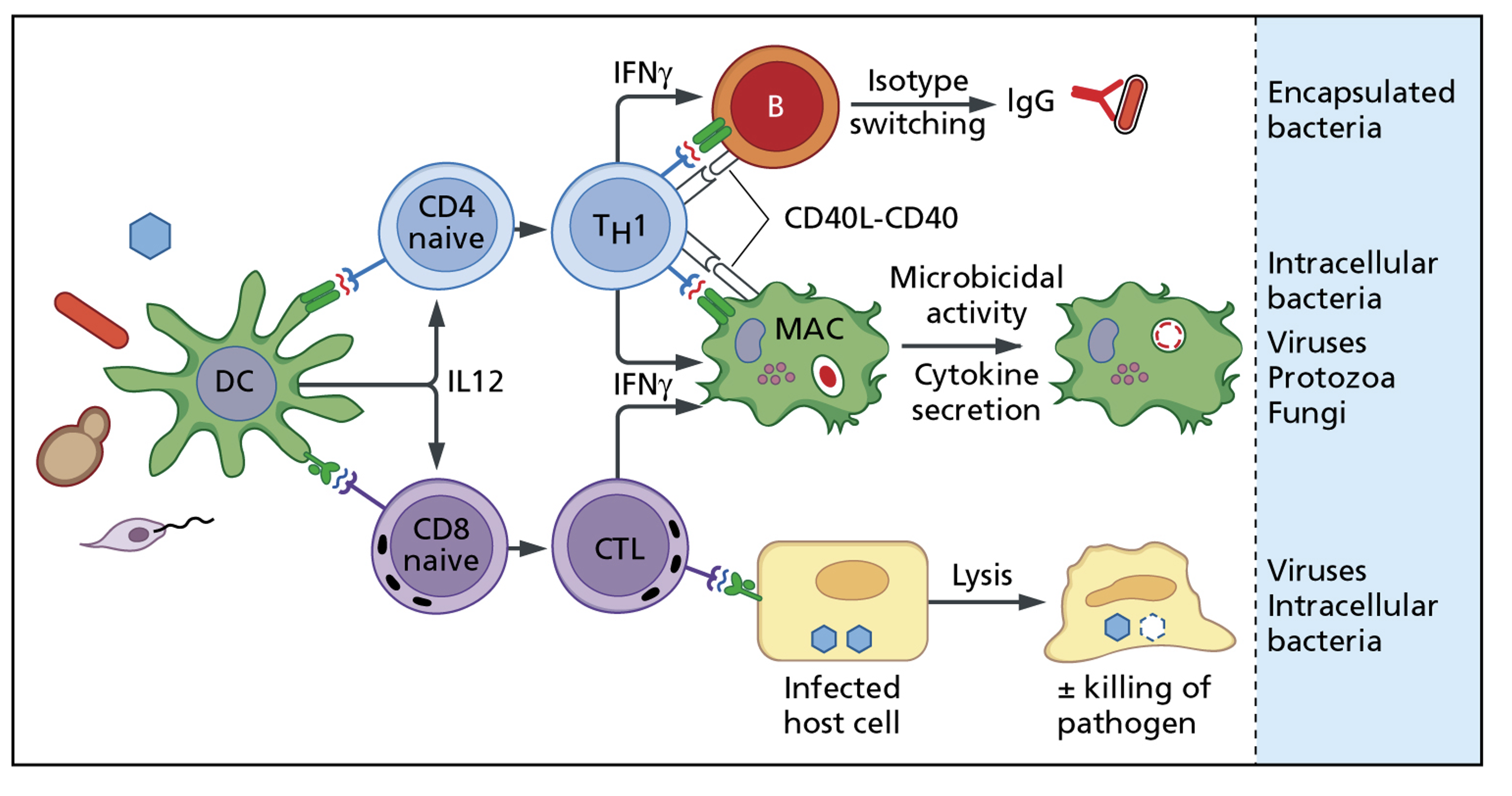
Cell-mediated immunity - Th1 pathway


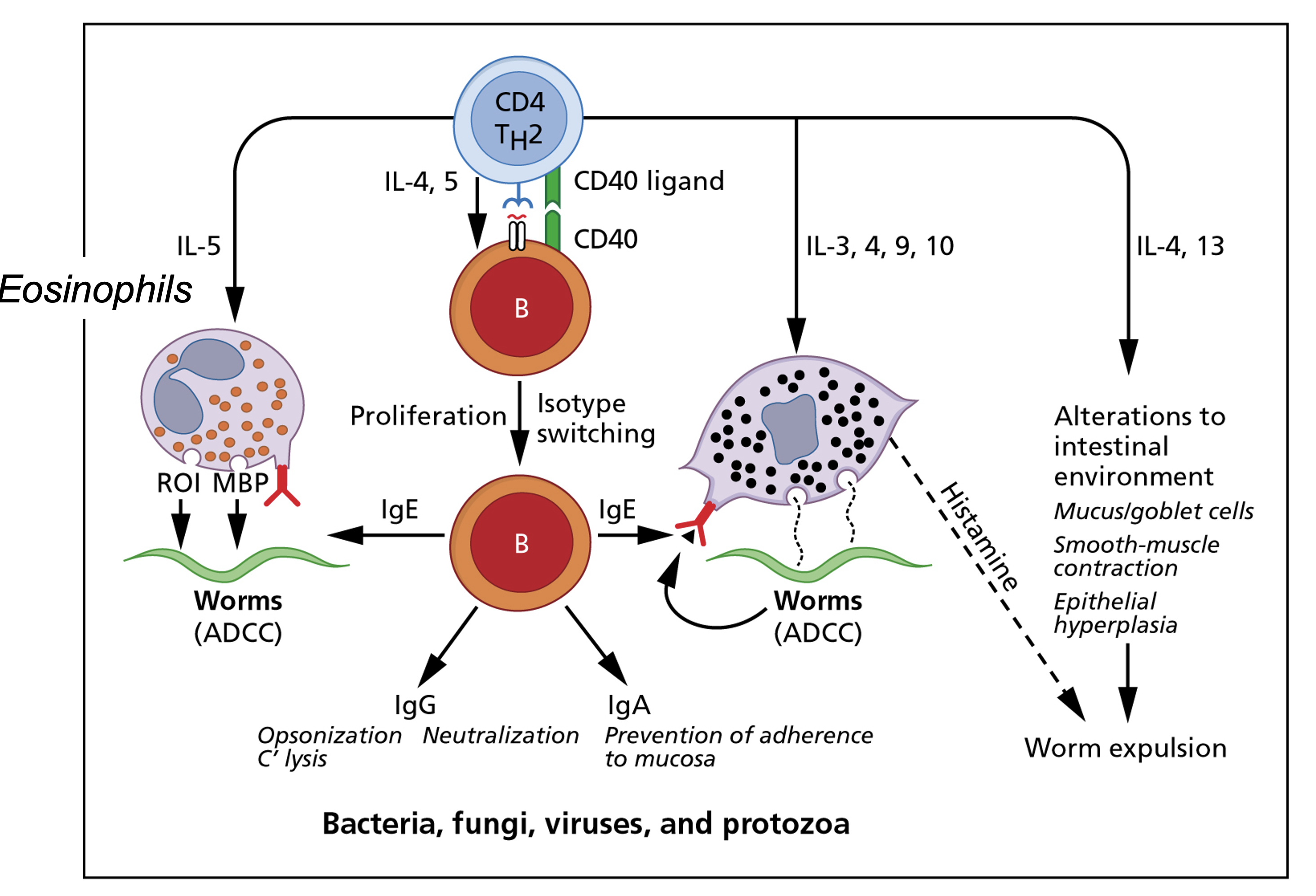
Cell-mediated immunity - Th2 pathway


Cell-mediated immunity -
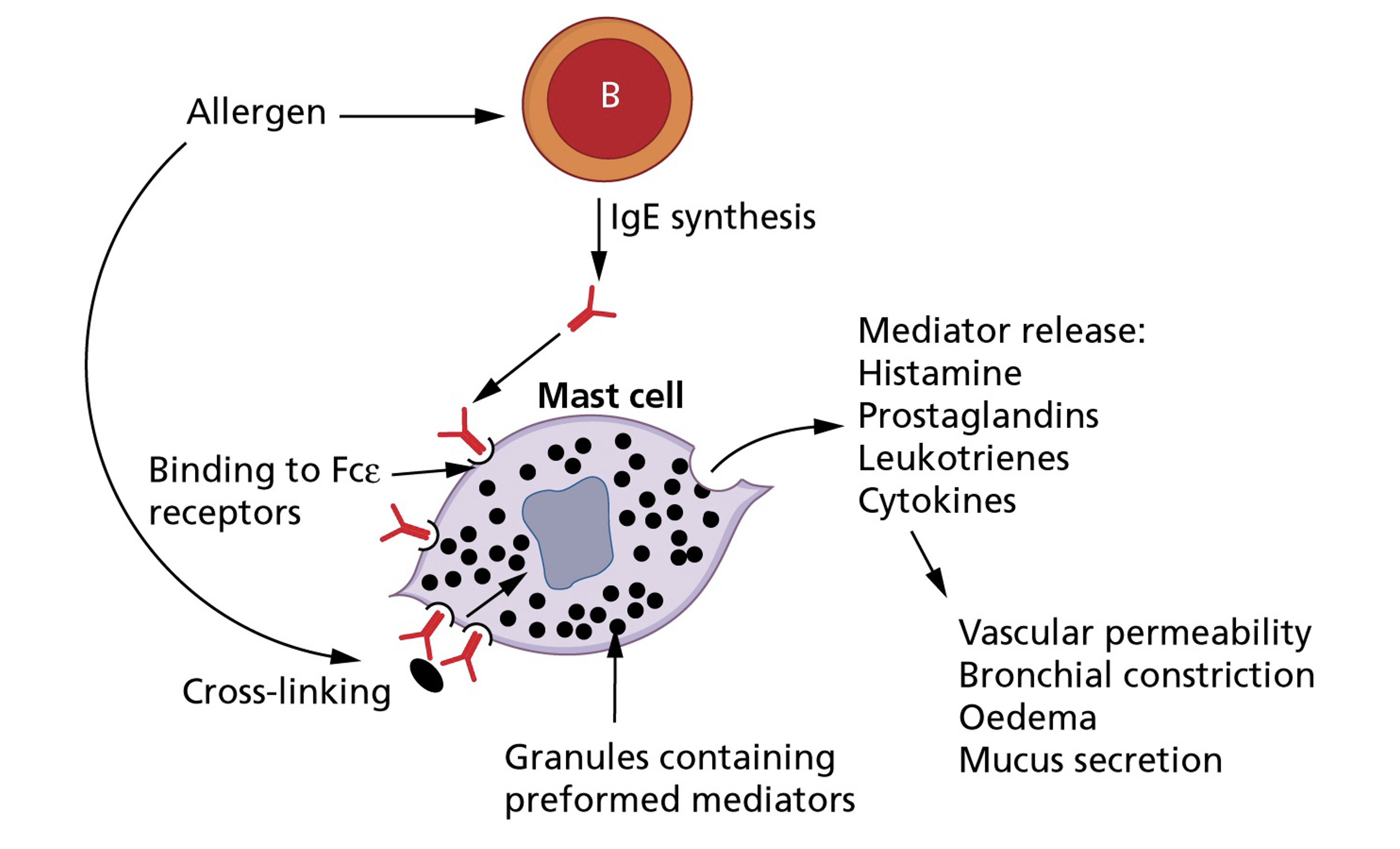
Drug allergy (Type IV hypersensitivity)

Infections in immunocompromised hosts:
A summary in one slide!


Etiology of fever in HIV-Associated FUO (n=70)

Etiology No. (%)
Infection 63 (88%)
 DMAC 22 (31%)
Pneumocystis jirovecii pneumonia 10 (13%)
 CMV 8 (11%)
 Histoplasmosis 5 (7%)
 Viral (not CMV) 5 (7%)
 Bacterial 4 (5%)
Mycobacterium tuberculosis 4 (5%)
 Fungal (not histoplasmosis) 2 (3%)
 Parasitic 2 (3%)
Mycobacterium genavense 1 (1%)
Neoplasia 6 (8%)
 Lymphoma 5 (7%)
 Kaposi sarcoma 1 (1%)
Miscellaneous 3 (4%)
 Drug fever 2 (3%)
 Castleman disease 1 (1%)

Naproxen (NSAID) fever suppression test for “tumor fever”


  • Indications for trial:
    • Temperature > 37.8°C at least once daily for ≥2 weeks
    • Lack of evidence of infection (physical exam, labs, imaging)
    • Absence of drug fever, transfusion reaction, or allergic mechanisms
    • Lack of response to ≥7 days of empiric antibiotics
  • Procedure:
    • Naproxen 500 mg BID × 3-5 days
  • Positive result:
    • Prompt complete defervescence (lysis of fever)
    • Sustained normal temperature while receiving naproxen
    • Fever recurrence when drug discontinued
  • Note: Not universally used; sensitivity/specificity debated

Diagnosis of FUO

General diagnostic evaluation of FUO


Step Investigation
History & Exam Comprehensive history; repeated physical exams
Laboratory CBC, comprehensive metabolic panel, urinalysis with microscopy
Inflammatory Markers ESR, C-reactive protein
Autoimmune Screening ANA, rheumatoid factor
Imaging - First Line Chest radiograph, CT abdomen/pelvis
Cultures Blood cultures (3 specimens without antimicrobials), urine culture
Serologies CMV IgM/PCR, heterophil antibody (EBV) in young adults
Tuberculosis Tuberculin skin test, interferon-gamma release assay
Advanced Imaging MRI, PET-CT, duplex ultrasound lower extremities
Invasive Procedures Biopsy (lymph node, liver, bone marrow) if indicated

Patient history


  • Helps guide choice of initial laboratory investigations - this is the most important step

  • Travel history: where, when, duration, exposures to animals, arthropod vectors, contaminated water

  • Exposure to animals and work environment: pet birds (psittacosis), cats (toxoplasmosis, bartonellosis), tick exposure (Lyme, Q fever)

  • Recent contact with ill persons or family history of FUO (e.g., familial Mediterranean fever)

  • Complete medication list: including OTC drugs, supplements, recent antimicrobials

  • Prior history of FUO - may be recurrence of same diagnosis

  • Previously diagnosed conditions: malignancy, rheumatic fever, valve disease that predisposes to endocarditis

Verification of fever and pattern of fever


  • Verification of fever: Often overlooked step
    • In some series, up to 30% referred for FUO where determined to NOT have fever
    • Request fever log or have patient take temperatures at home
  • Fever patterns - use of patterns to narrow differential:
    • Continuous, remittent, intermittent, hectic (Charcot’s), quotidian, quartan, biphasic (saddleback), Pel-Ebstein
  • Factors affecting fever pattern:
    • Hydration status, ambient temperature
    • Accuracy of temperature measurements (different sites)
    • Antipyretics and corticosteroids suppress fever
    • Blood transfusions and other medical interventions

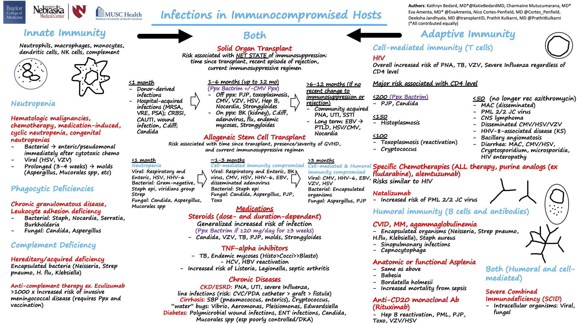
Continuous sustained fever


  • Continuous fever with slight remission not exceeding 2°C

    • Lobar and gram-negative pneumonia
    • Rickettsiosis
    • Typhoid fever
    • CNS disorders
    • Tularemia
    • Falciparum (malignant tertian) malaria

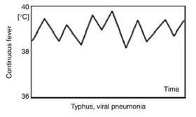
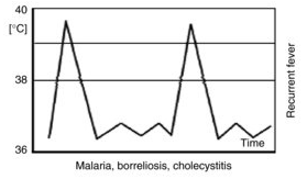
Intermittent (quotidian) fever


  • Intermittent fever with wide fluctuations, usually normal or low in the morning and peaking between 4:00 and 8:00 PM
    • Localized pyogenic infections and bacterial endocarditis with chills and leukocytosis
    • Malaria may present with daily (quotidian), every 3rd day (tertian), or every 4th day (quartan) patterns
    • Double quotidian pattern (two daily spikes) seen with:
      • Salmonellosis
      • Miliary tuberculosis
      • Double malarial infections (>1 species)
      • Gonococcal and meningococcal endocarditis

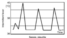
Malaria fever - Paroxysmal patterns

Febrile paroxysms may occur every other day for P. vivax, P. ovale, and P. falciparum (tertian fever) and every third day for P. malariae (quartan fever).

Paroxysms occurring at regular intervals are more common in P. vivax or P. ovale than P. falciparum. With improvements in early diagnosis and treatment, this traditional description of cyclic fever is seen infrequently.

Saddle-back (biphasic) fever


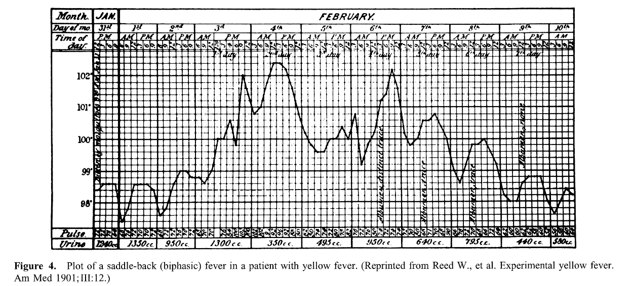
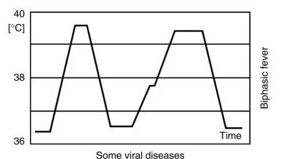
  • Several days of fever, distinct reduction in fever for ~1 day, then several days of higher fever
    • Dengue and yellow fever
    • Colorado tick fever
    • Rift valley fever
    • Influenza and other viral infections

Intermittent hectic (Charcot’s) fever


  • Sporadic episodes of fever with periods of normal temperature and recurrence
    • Frequently seen in cholangitis associated with cholelithiasis
    • Classic teaching: jaundice, fever, right upper quadrant pain (Charcot’s triad)
    • Often associated with leukocytosis and toxic appearance

Undulating (Pel-Ebstein) fever


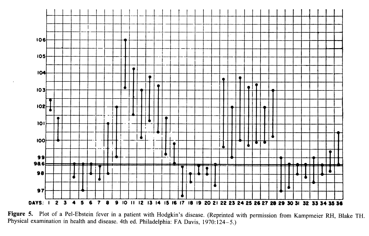
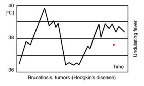
  • Weekly or longer periods of fever and equally long afebrile periods, with repetition of the cycle
    • Hodgkin’s disease (classic association)
    • Brucellosis due to Brucella melitensis
    • Occasionally tuberculosis

Typus Inversus


  • Reversal of diurnal pattern with highest temperatures in early morning hours rather than late afternoon/evening
    • Miliary TB
    • Salmonelloses
    • Hepatic abscess
    • Bacterial endocarditis

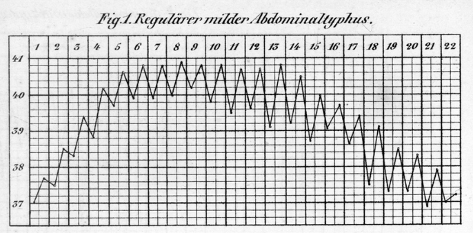
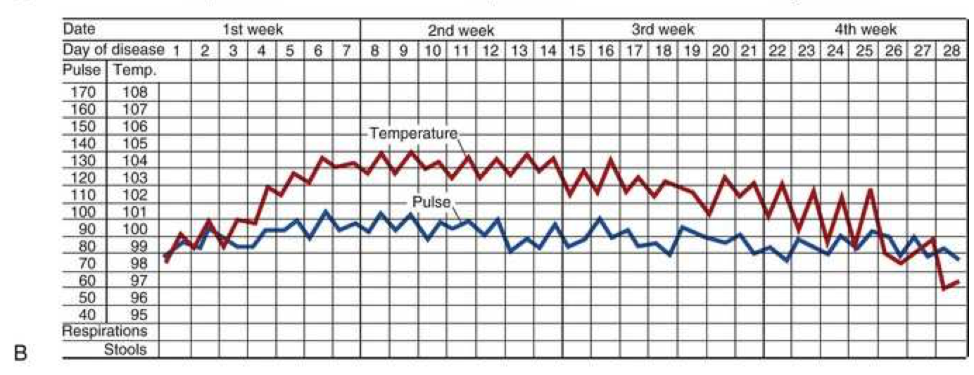
Typhoid fever - Step-ladder fever

Jarisch-Herxheimer reaction

  • Definition: Sharply increased elevation of temperature with rigors, chills, and constitutional symptoms occurring within hours of starting antibiotic therapy

  • Mechanism: Lysis of spirochetes (or other organisms) releases endotoxin triggering acute inflammatory response

  • Organisms associated:

    • Primary or secondary syphilis
    • Leptospirosis
    • Tick-borne relapsing fever (Borrelia)
    • Tetracycline or chloramphenicol therapy for acute brucellosis
  • Clinical significance: Can be severe enough to cause hemodynamic compromise; does not indicate treatment failure
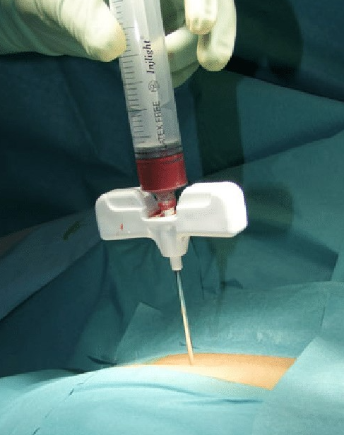
Physical examination


  • Some signs are subtle and may require repeated exams to be appreciated
  • Vigorous search for lymphadenopathy (consideration for biopsy)
  • Careful examination of fundi, oropharynx, temporal arteries, abdomen, spleen, joints, skin, nails, genitalia
Body site Physical finding Diagnosis
Head Sinus tenderness Sinusitis
Temporal artery Nodules, reduced pulsations Temporal arteritis
Oropharynx Ulceration; tender tooth Disseminated histoplasmosis, periapical abscess
Fundi / conjunctivae Choroid tubercle, petechiae, Roth’s spot Disseminated granulomatosis, endocarditis
Thyroid Enlargement, tenderness Thyroiditis
Heart Murmur Infective or marantic endocarditis
Abdomen Enlarged iliac lymph nodes, splenomegaly Lymphoma, endocarditis, disseminated granulomatosis
Rectum Perirectal / prostatic fluctuance, tenderness Abscess
Genitalia Testicular nodule; epididymal nodule Periarteritis nodosa; disseminated granulomatosis
Lower extremities Deep venous tenderness Thrombosis / thrombophlebitis
Skin and nails Petechiae, splinter hemorrhages, subcutaneous nodules, clubbing Vasculitis, endocarditis

Laboratory investigations


“The cause of FUO is more frequently a common disease presenting in an atypical fashion than a rare disease presenting in a typical fashion.


  • Multiple diagnostic algorithms exist in literature

  • Must be selectively applied or will result in excessive unfocused diagnostic testing

    • False positives lead to misdiagnosis
    • Misguided treatment plans
  • “Sutton’s Law” - pursue most likely diagnosis first based on history and epidemiology

  • History and physical exam (most important) should guide choice and sequence of tests

Examples of potential diagnostic clues (1/3)


Etiology Historical clues Physical clues
Anaplasmosis Ixodes tick bite; outdoor activity in North Central / Eastern US Fever, headache, arthralgia, myalgia, pneumonitis, thrombocytopenia, lymphopenia, ↑ liver enzymes
Babesiosis Ixodes tick bite; outdoor activity in Northeastern US Arthralgias, myalgias, relative bradycardia, hepatosplenomegaly, anemia, thrombocytopenia, ↑ liver enzymes
Bartonellosis Travel to Andes (Oroya fever; B. bacilliformis); homelessness (B. quintana); scratch from infected kitten/cat (B. henselae) Conjunctivitis, retro-orbital pain, anterior tibial bone pain, macular rash, nodular plaque lesions, regional lymphadenopathy
Blastomycosis Contact with soil near Mississippi/Ohio River valleys, Saint Lawrence River, or Great Lakes; exposure to infected dogs Arthritis, atypical pneumonia, pulmonary nodules, ARDS, verrucous/nodular/ulcerative skin lesions, prostatitis
Brucellosis Contact with/consumption of products from infected goats, pigs, camels, yaks, buffalo, cows; abattoir work Arthralgias, hepatosplenomegaly, suppurative musculoskeletal lesions, sacroiliitis, spondylitis, uveitis, hepatitis, pancytopenia
Coccidioidomycosis Exposure to soil or dust in the southwestern US Arthralgias, pneumonia, pulmonary cavities, pulmonary nodules, erythema multiforme, erythema nodosum
Ehrlichiosis Amblyomma, Dermacentor, or Ixodes tick bite; outdoor activity in midwestern / southeastern US Pneumonitis, hepatitis, thrombocytopenia, lymphopenia

Examples of potential diagnostic clues (2/3)


Etiology Historical clues Physical clues
Enteric fever (Salmonella Typhi) Recent travel to endemic country; consumption of potentially contaminated food or water Headache, arthritis, abdominal pain, relative bradycardia, hepatosplenomegaly, leukopenia
Histoplasmosis Exposure to bat/blackbird excreta in roosts, chicken houses, or caves; Ohio and Mississippi River valleys Headache, pneumonia, pulmonary cavities, mucosal ulcers, adenopathy, erythema nodosum, erythema multiforme, hepatitis, anemia, leukopenia, thrombocytopenia
Leptospirosis Occupational exposure in sewers, rice/sugar cane fields, abattoirs; recreational water sports; contact with contaminated water or infected dogs Bitemporal/frontal headache, calf and lumbar muscle tenderness, conjunctival suffusion, hepatic and renal failure, hemorrhagic pneumonitis
Leishmaniasis (visceral) Recent travel to sand fly–endemic areas Hepatosplenomegaly, lymphadenopathy, hyperpigmentation of face/hands/feet/abdomen (kala azar)
Malaria Recent travel to endemic areas in Asia, Africa, or Central/South America Fever, headaches, nausea, vomiting, diarrhea, hepatosplenomegaly, anemia
Psittacosis (Chlamydia psittaci) Contact with birds, especially psittacine birds Fever, pharyngitis, hepatosplenomegaly, pneumonia, blanching maculopapular eruptions, erythema multiforme, erythema marginatum, erythema nodosum

Examples of potential diagnostic clues (3/3)


Etiology Historical clues Physical clues
Q fever (Coxiella burnetii) Farm, veterinary, or abattoir work; unpasteurized milk; contact with infected sheep, goats, or cattle Atypical pneumonia, hepatitis, hepatomegaly, relative bradycardia, splenomegaly
Rat-bite fever (Streptobacillus moniliformis) Recent bite/scratch from rat, mouse, or squirrel; ingestion of food/water contaminated with rat excrement Headaches, myalgias, polyarthritis, maculopapular/morbilliform/petechial/vesicular/pustular rash over palms, soles, and extremities
Relapsing fever (Borrelia recurrentis) Poverty/crowding/poor sanitation (louse-borne); camping in the Grand Canyon (tick-borne) High fever with rigors, headache, delirium, arthralgias, myalgias, hepatosplenomegaly
Rocky Mountain spotted fever Outdoor activity in South Atlantic or southeastern US; Dermacentor tick bites Headache, petechial rash on extremities, palms, and soles
Tuberculosis Contact with TB; immigration from endemic country; homeless shelter or healthcare facility exposure Night sweats, weight loss, atypical pneumonia, cavitary pulmonary lesions
Tularemia Bites from Amblyomma or Dermacentor ticks, deer flies, or mosquitoes; direct contact with rabbits, squirrels, deer, raccoons, cattle, sheep, or swine Ulcerated skin lesion at bite site, pneumonia, relative bradycardia, lymphadenopathy, conjunctivitis
Whipple’s disease (Tropheryma whipplei) Potential association with exposure to sewage Chronic diarrhea, arthralgia, weight loss, malabsorption, malnutrition

Bone marrow biopsy


  • Diagnostic yield: ~25% in two case series; especially valuable for:
    • Granulomatous infections (TB, histoplasmosis, sarcoidosis)
    • Hematologic malignancies (leukemia, lymphoma)
    • Patients with abnormal CBC (anemia, thrombocytopenia, leukopenia)
  • Organisms identified:
    • Mycobacterium tuberculosis
    • Fungi (Histoplasma, Cryptococcus, Coccidioides)
    • Intracellular bacteria (Brucella, Bartonella)
  • Consider: When fever pattern and presentation suggest granulomatous disease or hematologic malignancy


Imaging studies


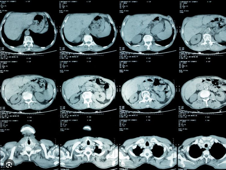
Generally low diagnostic yield without localizing symptoms

  • CT of abdomen and chest

  • Ultrasound of gallbladder and hepatobiliary systems

  • CT pulmonary angiogram (for pulmonary embolis)

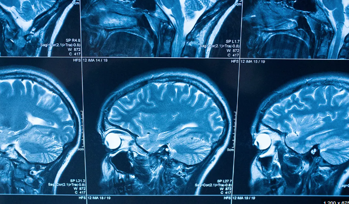
  • MRI for CNS, spleen, lymph nodes, aorta (vasculitis)

  • Indium 111-tagged white blood cell scan (becoming less common)

  • Gallium-67 scan (largely replaced by PET-CT)

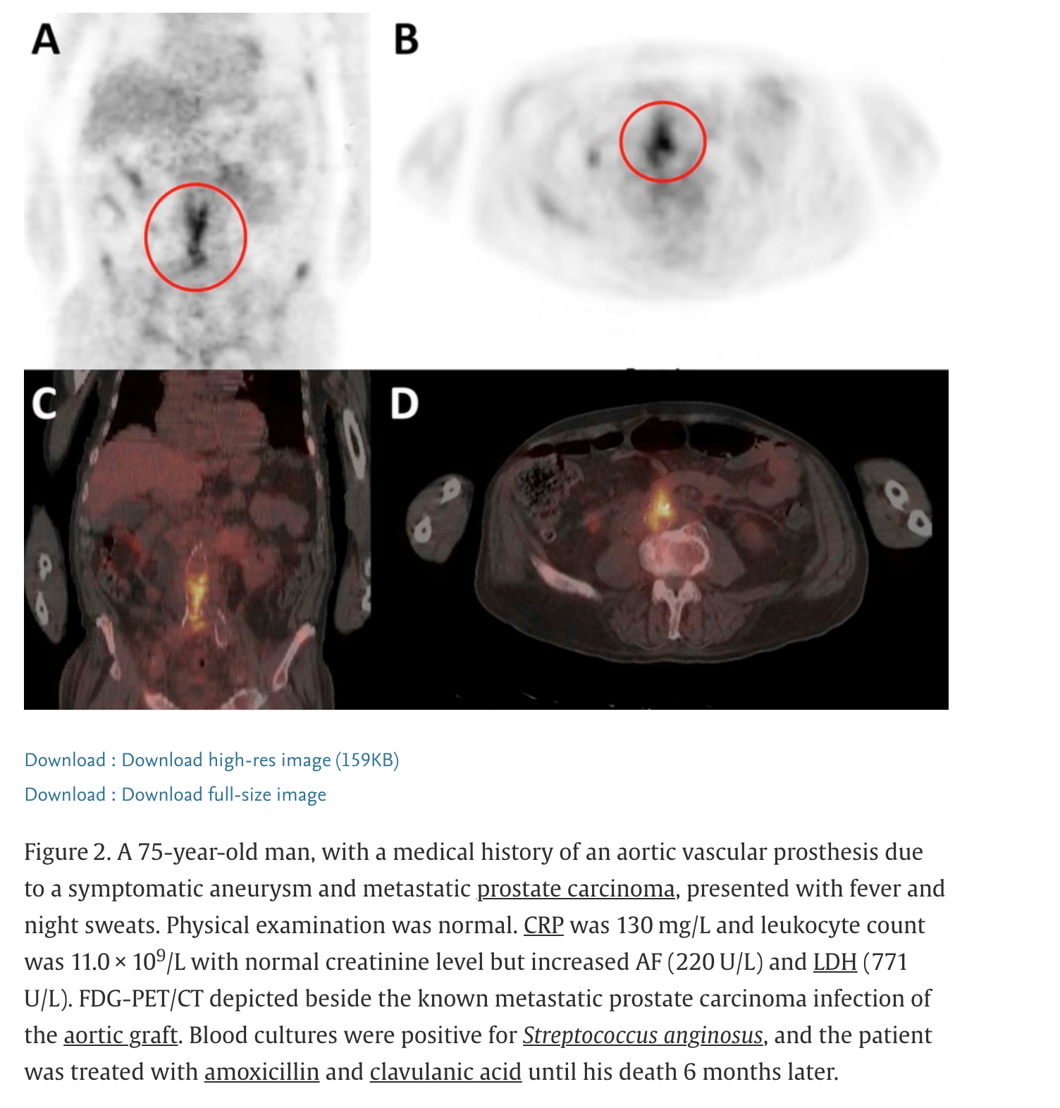
  • PET-CT: Superior sensitivity for inflammatory and malignant processes

¹⁸F-fluorodeoxyglucose (FDG) positron emission tomography


Invasive diagnostic procedures


  • Biopsy techniques:
    • Excisional biopsy (lymph node)
    • Needle biopsy (liver, kidney, muscle)
    • Laparotomy or laparoscopy for direct visualization
  • Diagnostic yield:
    • Biopsy yields diagnosis in < 50% of cases
    • Average 2-3 biopsies needed to establish diagnosis
  • Indications:
    • Abnormal imaging or physical exam findings
    • Lymphadenopathy suitable for lymph node biopsy
    • Suspected granulomatous disease
    • Persistent fever despite extensive workup

Treatment Section


A fundamental principle in classic FUO:

Therapy should be withheld until the cause of fever is determined

Diagnostic summary and approach

Step 1
Perform a comprehensive history and complete physical examination.
Step 2
PDCs identified?
YES →
NO
Step 3
Order CBC, CMP, ESR, CRP, ferritin, TSH, RF, ANA, HIV-1/2 serology, hepatitis A, B, and E, urinalysis with microscopy, tuberculosis skin test or TB whole-blood interferon-γ releasing assay, blood cultures ×3, venous duplex imaging of the lower extremities, CT of chest and abdomen/pelvis, and echocardiography (TTE/TEE).
Step 4
Any positive first-tier testing?
YES →
NO
Step 5
Any PDCs by repeat history and physical examination?
YES →
NO
Step 6
Order 18FDG-PET/CT scan imaging (if not already performed).
Positive →
Negative Result
Step 7
If there are still no new PDCs, re-evaluate the patient history and physical examination periodically. Continue to monitor for any new PDCs and apply appropriate noninvasive and/or invasive diagnostic testing based upon any new findings. At this stage, empiric use of nonsteroidal anti-inflammatory agents or immunosuppressant therapy such as corticosteroids could be entertained after discussing risks and benefits with the patient.
Step 2 — If YES
Apply appropriate invasive or noninvasive diagnostic studies for investigation.
Step 4 — If YES
Apply second-tier testing such as specialized imaging (MRI, nuclear imaging, or 18FDG-PET scans) or appropriate biopsy methods for culture and histologic analysis.
Step 5 — If YES
Apply appropriate invasive and noninvasive diagnostic studies for investigation.
Step 6 — Positive Result
Apply appropriate invasive and noninvasive diagnostic studies for investigation.
Adapted from Cunha et al. © 2020 JHU/AAM

When is immediate treatment indicated?


  • Suspected temporal arteritis
    • Empirical corticosteroids to prevent vascular complications (blindness, stroke)
    • Do not wait for biopsy confirmation if clinical suspicion high
  • Febrile neutropenia or severe immunocompromise
    • High prevalence of serious bacterial infections
    • Broad-spectrum antimicrobial therapy with anti-pseudomonas coverage after appropriate cultures obtained
    • No delay for culture results
  • Select cases with strong clinical suspicion:
    • Anti-mycobacterial therapy in suspected TB (especially if respiratory symptoms)
    • Targeted therapy based on epidemiology and presentation

Prognosis

  • Determined by: The underlying cause of fever and nature of underlying disease(s)

  • Poor prognosis: Elderly patients with malignant neoplasms

  • Diagnostic delay worsens prognosis in:

    • Intra-abdominal infections (perforation, sepsis)
    • Miliary tuberculosis
    • Disseminated fungal infections
    • Recurrent pulmonary emboli
  • Favorable outlook: Patients with undiagnosed FUO after extensive evaluation

    • Most experience resolution of fever within 4 weeks without sequelae
    • 5-year mortality rate: 3.2% for undiagnosed FUO
    • Superior to patients with diagnosed malignancy as the underlying cause


References


Corey L, Boeckh M. Persistent fever in patients with neutropenia. New England Journal of Medicine 2002;346:222–4.
Gabay C, Kushner I. Acute-phase proteins and other systemic responses to inflammation. New England Journal of Medicine 1999;340:448–54.
Haidar G, Singh N. Fever of Unknown Origin. New England Journal of Medicine 2022;386:463–77.
Kouijzer IJE, Vos FJ, Rijnders BJA, Aarntzen EAHG, Bleeker-Rovers CP. The value of \(^{18}\)F-FDG PET/CT in diagnosing infectious endocarditis. European Journal of Nuclear Medicine and Molecular Imaging 2018;45:1085–99.
Mackowiak PA, Wasserman SS, Levine MM. Concepts of fever: Recent advances and lingering dogma. Clinical Infectious Diseases 1997;25:996–1003.
Mackowiak PA, Worden G. Carl Reinhold August Wunderlich and the evolution of clinical thermometry. Clinical Infectious Diseases 1994;18:458–67.
Playfair JHL, Bancroft GJ. Infection and Immunity. 4th ed. Oxford University Press; 2013.
Sajadi MM, Mackowiak PA. Chapter 57: Temperature Regulation and the Pathogenesis of Fever. Mandell’s principles and practice of infectious diseases. 9th ed., Elsevier; 2020.
Sickles EA, Young VM, Greene WH, Wiernik PH. Clinical presentation of infection in granulocytopenic patients. Archives of Internal Medicine 1975;135:715–9.
Wright WF, Auwaerter PG. Chapter 58: Fever of Unknown Origin. Mandell’s principles and practice of infectious diseases. 9th ed., Elsevier; 2020.
Wright WF, Mackowiak PA. Fever of Unknown Origin. Mandell’s principles and practice of infectious diseases. 8th ed., Elsevier; 2015.